Middle Ear Effusion
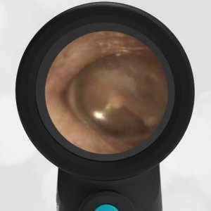
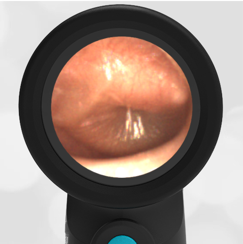
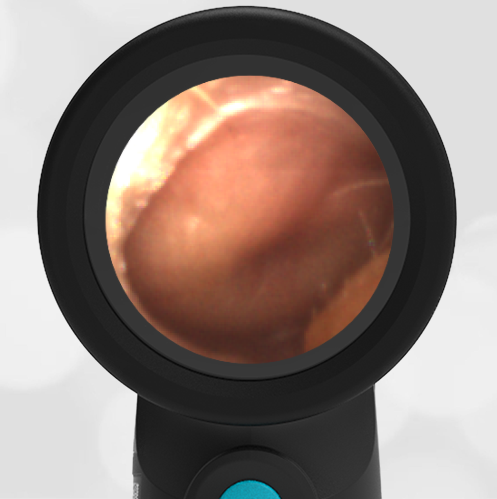
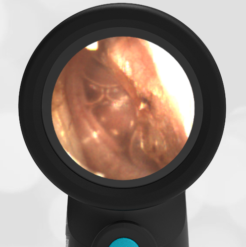
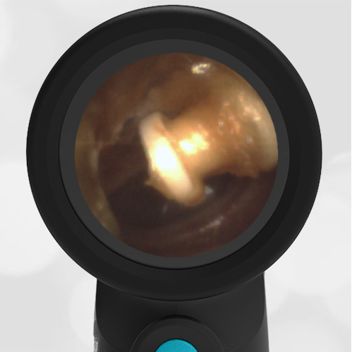
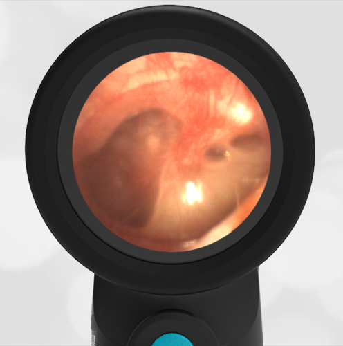
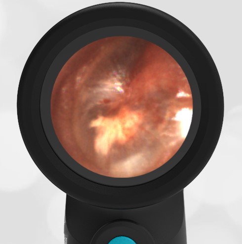
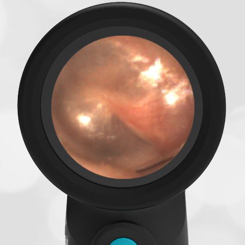
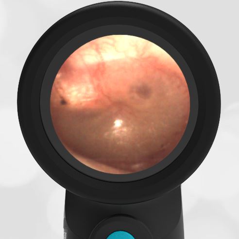
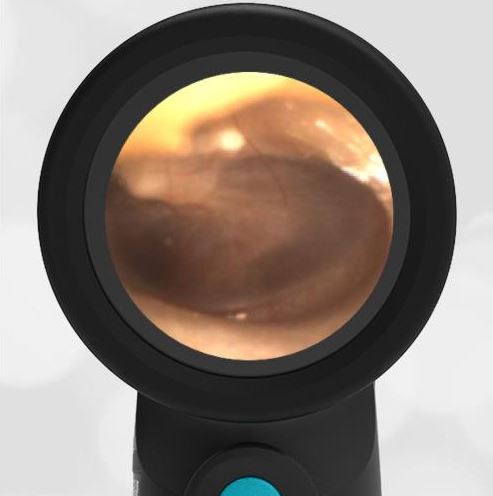
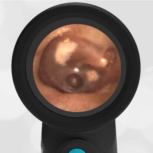
An 8-year-old female with a history of ear infections presents to the pediatric clinic for a well-child check. She has had mild cold-like symptoms including rhinorrhea and congestion. She does not have a fever, and she does not complain of ear pain. She continues to eat and drink well and be active. Examination of the right ear reveals this image.
The child has a middle ear effusion (MEE).
This is a common clinical scenario. A previously well child with mild viral symptoms. The ear shows air-fluid levels consistent with a middle ear effusion (MEE, fluid in the middle ear space, behind the tympanic membrane). The air-fluid levels in the image are at the 6 o’clock position. In reality, because of the way the Wispr otoscope is being held, the air-fluid levels are anatomically in the anterior-superior region. The presence of air along with fluid in the middle ear space suggests that the eustachian tube is functioning properly. Because the eustachian tube is functioning properly to equalize the pressure on both sides of the eardrum, there is no bulging as would be seen in acute otitis media (AOM). In addition, there is no indication for intervention such as antibiotics. The fluid will drain through the eustachian tube with time.
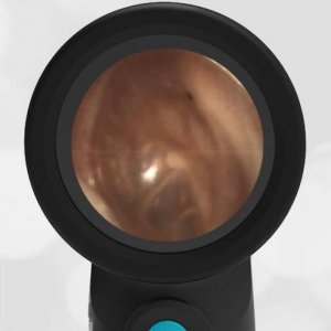
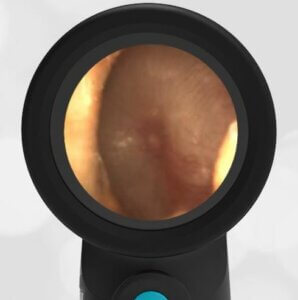
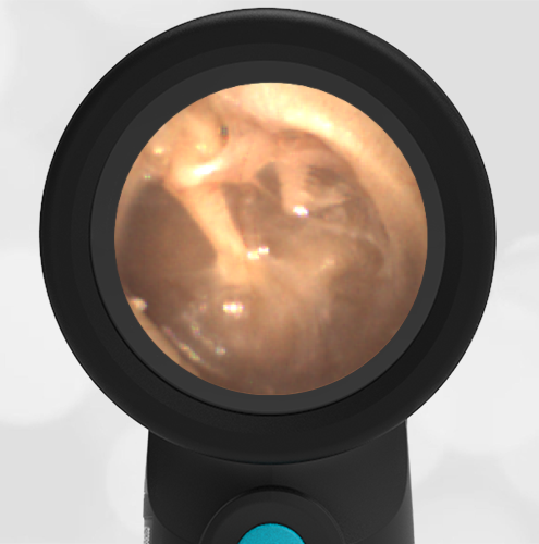
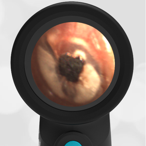
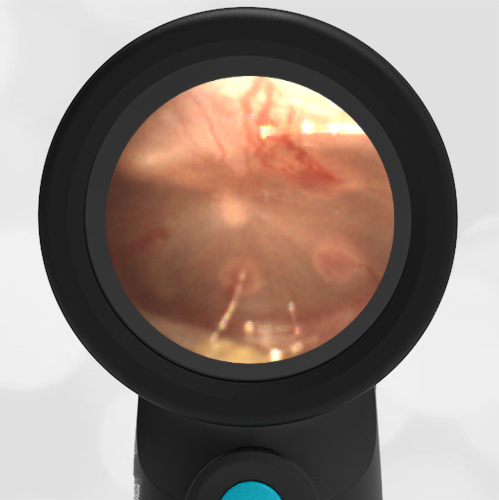
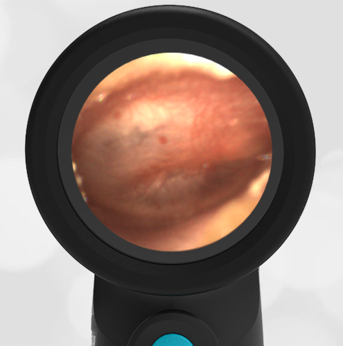
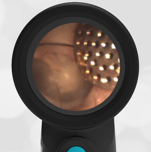
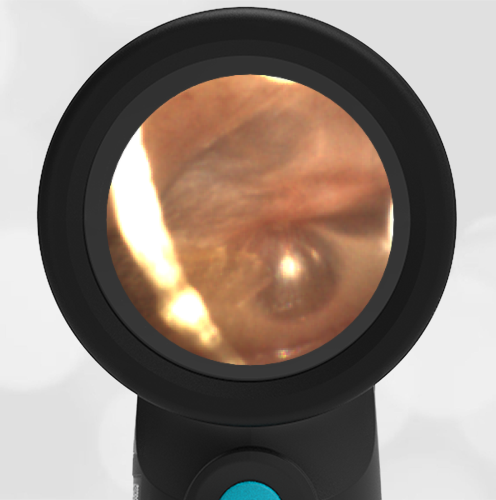
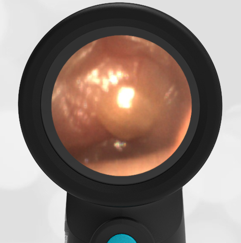
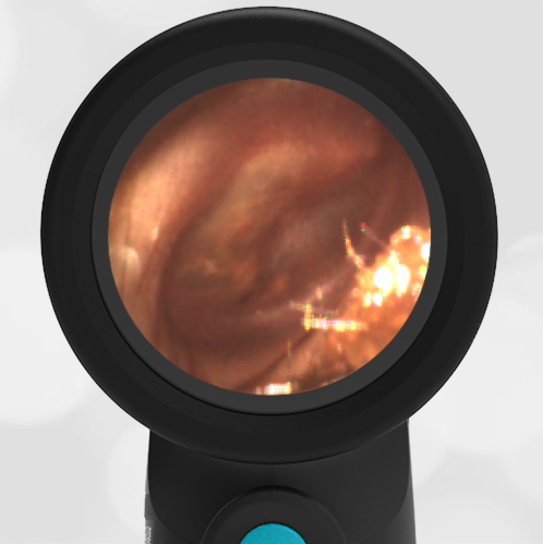
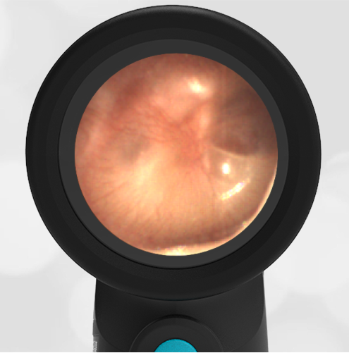
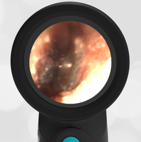
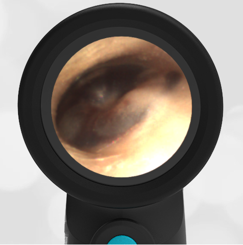
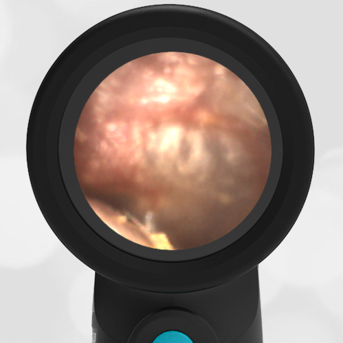
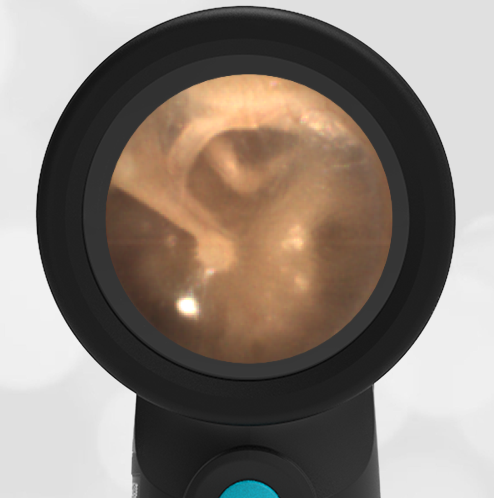
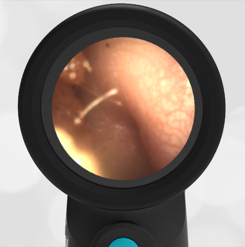
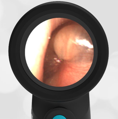
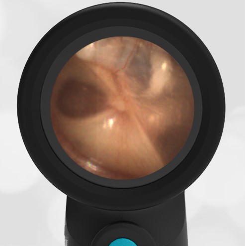
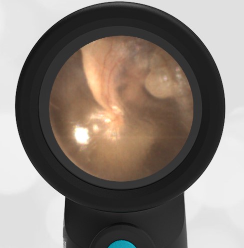
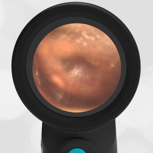
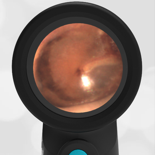
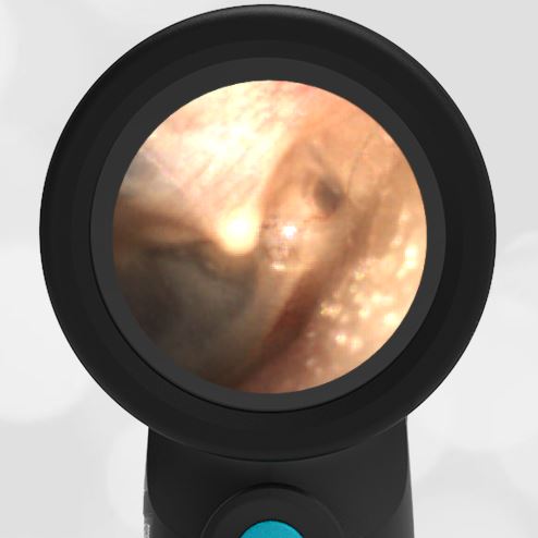
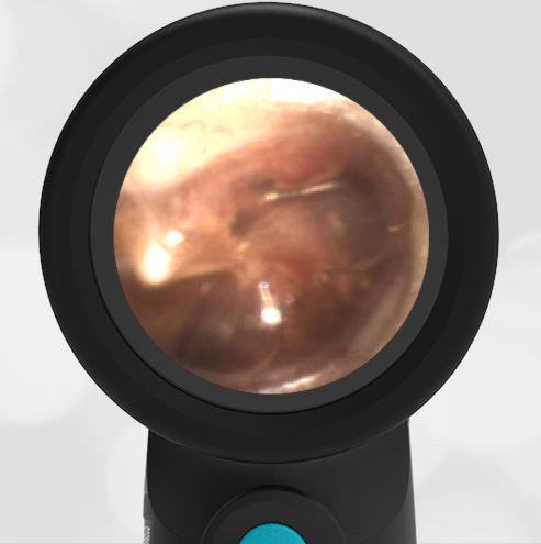
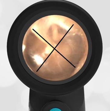
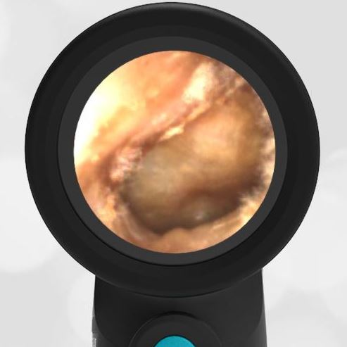
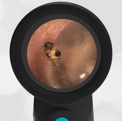
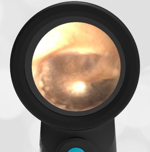
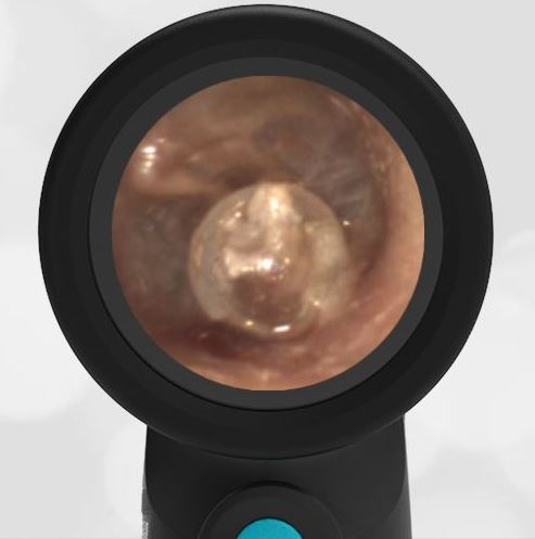
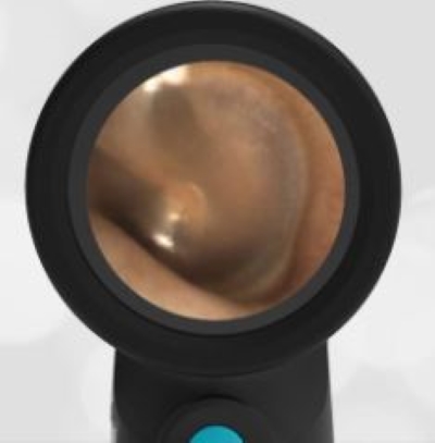
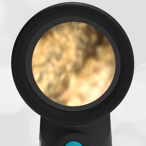
Compare the following images of eardrums: normal, middle ear effusion, and acute otitis media. You can clearly appreciate the difference between the MEE with a functioning eustachian tube and the AOM with profound bulging indicating a non-functioning eustachian tube.
- Normal Ear
- Middle Ear Effusion
- Acute Otitis Media (AOM)

