Pars Flaccida Retraction
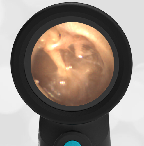
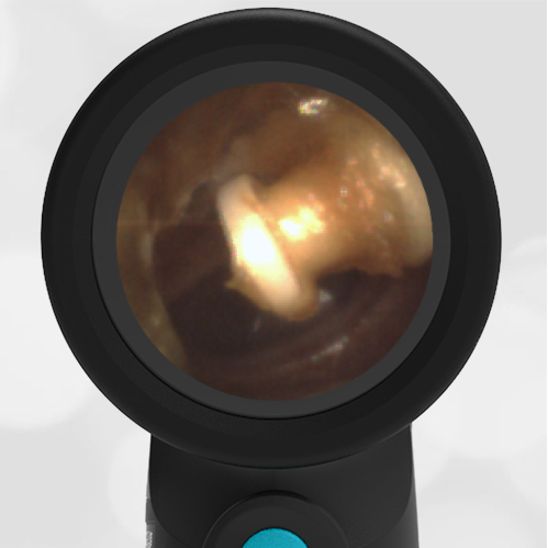
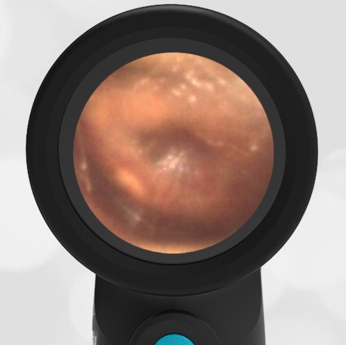
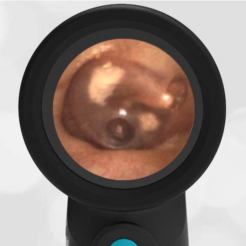
A 55 year old woman with a history of decreased hearing in her right ear presents for a routine physical exam. She has no new hearing complaints, although she affirms that she has a long history of poor hearing. Examination of her right ear reveals this image.
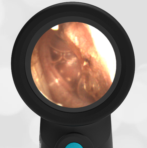
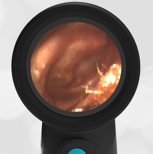
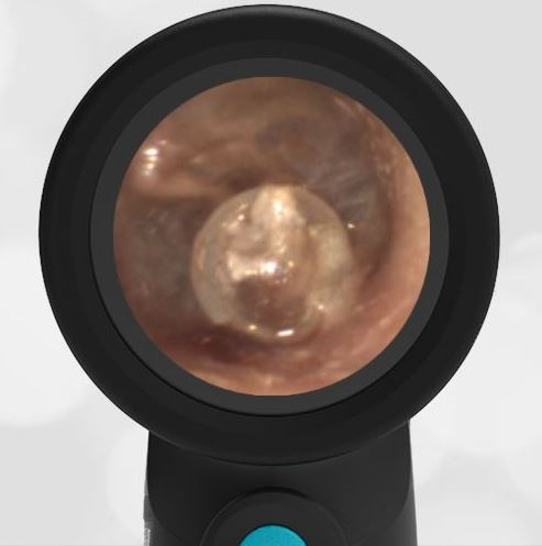
The patient has retraction and likely erosion of the portion of her tympanic membrane (eardrum) called the pars flaccida.
The pars flaccida is the superior portion of the eardrum and covers portions of the malleus and incus bones. In this image, abnormal anatomy of the pars flaccida is clearly visible with portions of the malleus and incus bones exposed. The portion of the eardrum responsible for the translation of sound waves to mechanical motion, the pars tensa, is intact. The poor hearing is likely due to damage to the articulating surfaces of the malleus, incus, and stapes bones.

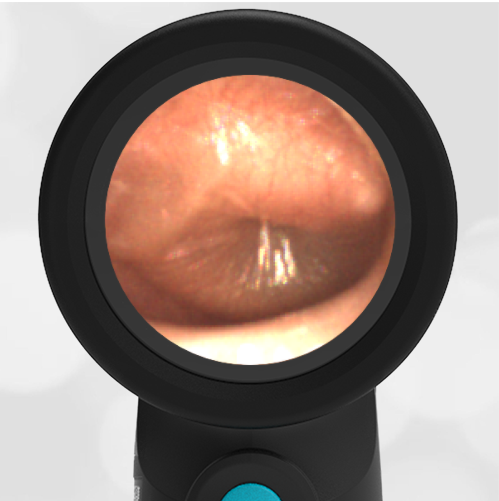
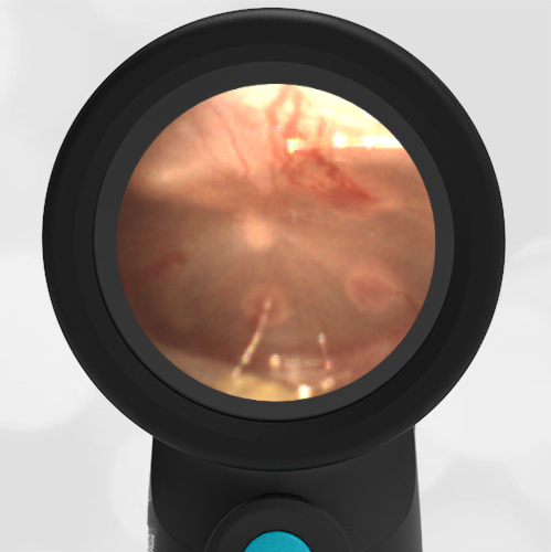
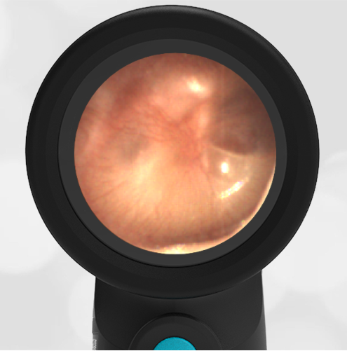
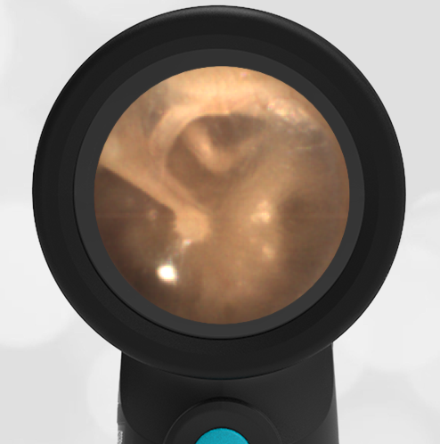
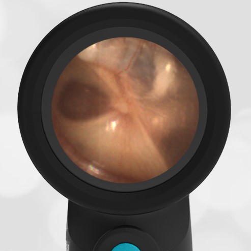
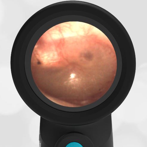
Compare the image from this patient with an image of a normal and healthy eardrum. The difference in the pars flaccida is apparent.
-
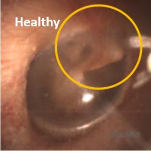
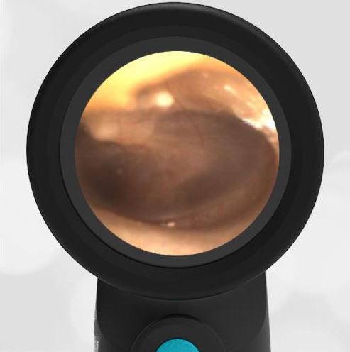
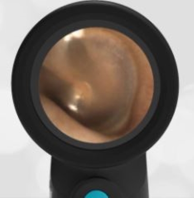
- Pars Flaccida – Healthy
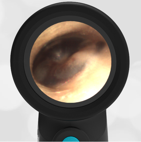
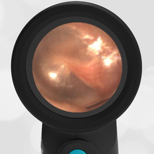
-
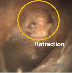
- Pars Flaccida – Retraction
If you would like to know more about ear anatomy, please see the WiscMed presentation on normal ear anatomy. It is available at Wispr University.