It can be difficult to distinguish between a normal ear and an ear with otitis media with effusion (OME). The hallmark of OME, air-fluid levels in the middle ear space, can be subtle. In this Wispr University article, we present a technique for making the air-fluid level more obvious.
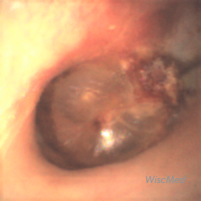
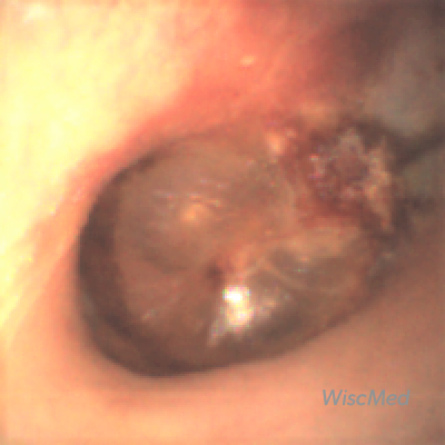
A 37 year-old woman returned from Hawaii complaining of right ear discomfort. While she was in Hawaii, she had viral symptoms including a sore throat, mild fever, cough and fatigue. She then developed right ear pain. She was seen at an urgent care clinic on the island and diagnosed with “an infection” along with an ear perforation and placed on an oral antibiotic and a topical ear antibiotic. Upon return to the mainland, the following image of her right ear was obtained with the Wispr digital otoscope. What is your diagnosis and proposed treatment at this point?
- Otitis Media with Effusion
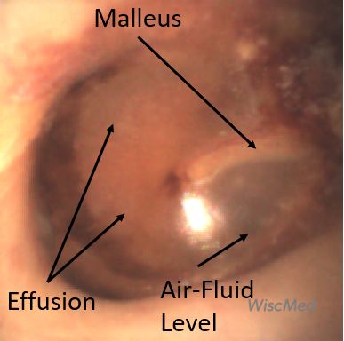
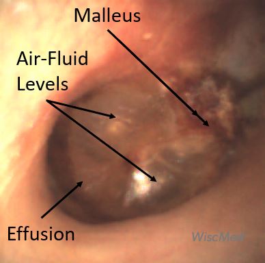
- Annotated
The patient has otitis media with effusion (OME). There is no perforation present. Antibiotics are not indicated solely for this condition.
One of the key advantages of digital otoscopy is the ability to carefully review exam findings “on the screen” after the exam has been completed. In this case, the key clinical feature is the air-fluid level associated with OME. It appears as a bubble behind the ear drum. It is a subtle finding. In addition, there is no bulging of the ear drum and the malleus is easily distinguishable. This rules out acute otitis media (AOM). There is no evidence of an ear drum perforation. The air-fluid levels indicate the Eustachian tube is functioning.
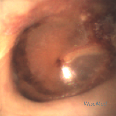
A middle ear effusion is fluid in the middle ear space, usually from inflammation secondary to a viral infection. The middle ear space is directly behind the ear drum. It’s partially visible to us because the ear drum is semi-transparent. After the initial exam, the patient was asked to perform the Valsalva maneuver. The Valsalva maneuver is pressure breath holding, familiar to us as we use the maneuver to “pop” our ears in a descending airplane. Here is the video of the patient performing the Valsalva maneuver. Note how obvious the air-fluid levels become as the increased air pressure in the middle ear moves the fluid about.
Complete exam video
And here is the post Valsalva image where the air-fluid levels are more distinct.
An interesting conclusion to this case is that when the presence of a OME is uncertain, having a cooperative patient perform the Valsalva maneuver may clarify the condition.

