2024 Best Digital Otoscopes Review
Dr. James Berbee, Founder of WiscMed
The promise of a digital otoscope is more reliable access to the tympanic membrane, improved patient comfort, medical documentation and sharing of the image with the patient, learners, parents, and colleagues.
There are many choices available for digital otoscopes. In this article, we compare six popular digital otoscopes.
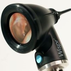
1. WiscMed Wispr Digital Otoscope, www.wiscmed.com
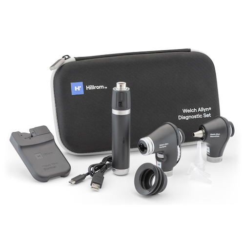
2. Welch Allyn I Examiner, https://www.hillrom.com/en/products/macroview-plus-otoscope/
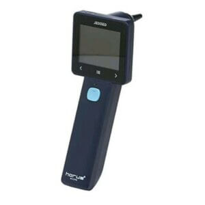
3. Jedmed Horus+ HD Video Otoscope, www.jedmed.com
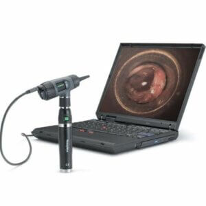
4. Welch Allyn Digital MacroView, www.hillrom.com/en/products/digital-macroview-otoscope/
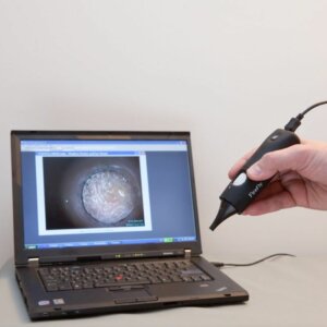
5. Firefly DE500 Video Otoscope, www.fireflyglobal.com
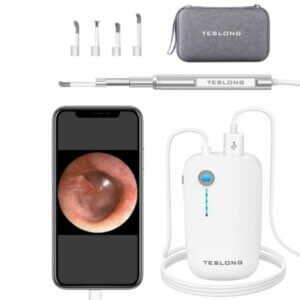
6. Teslong Digital Otoscope, www. amazon.com
We focus on a selection of devices that could be considered for use in the clinical environment. Each device has strengths and weaknesses. Which digital otoscope best serves your needs will depend on how you plan to use the device. All digital otoscopes have a camera, illumination, and some method for displaying an image. How they implement those components is quite different.
From the author of this document, Dr. James Berbee: I am an emergency physician, an inventor of the Wispr otoscope and the founder of the WiscMed company. This position prevents me from being the impartial reviewer that one would ideally prefer. However, I have attempted to be fair in my review, including pointing out the areas where the Wispr is not as strong as the other devices. If you believe I have made material mistakes in this document, please contact me. My contact information is at the end.
Digital Otoscope Comparison Chart
|
|
WiscMed Wispr | Welch Allyn I Examiner | Jedmed Horus+ | Welch Allyn Digital MacroView | Firefly DE500 | Teslong | |
 |
 |
 |
 |
 |
 |
||
| Cost | $1195 | $795 | $795 | $1150 | $319 | $59 | |
| Speculum Cost | $0.16/each | $0.05/each | $0.05/each | $0.05/each | $0.20/each | $0.27/each | |
Display and Image |
|||||||
| Display | LCD Built-in | Attaches to smartphone | LCD Built-in | Attaches to computer via USB | Attaches to computer via USB. Wireless version also available. | Attaches to smartphone. | |
| Screen Shape | Round | External Smartphone | Square | External Computer | External Computer | External Smartphone | |
| Touch Screen | Yes | External Smartphone | No, touch panel below the screen | External Computer | External Computer | External Smartphone | |
| Screen Size | 35 mm | External Smartphone | 50 mm x 40 mm | External Computer | External Computer | External Smartphone | |
| Camera location1 | Distal | Proximal | Proximal | Proximal | Proximal | Proximal | |
| Image Resolution | 249 x 250 | External Smartphone | 1440 x 1280 | 1280 x 1024 | 1280 x 1024 | 1280 x 720 | |
| Image Type | bmp, avi | Jpeg, MP4 | Jpeg, H.264 | Png, wmv | Bmp, avi | Jpeg, MP4 | |
| Size of image file | 0.5 MB | 230 KB | 0.5 MB | 1.5 MB | 3.8 MB | 27 KB | |
| Image Transfer | Via USB port to thumb drive | Via Smartphone | Via USB cable to computer | Via USB cable to computer | Via USB cable to computer | Via cables and interface box to smartphone | |
| Video Mode | Yes | Yes – Default | Yes | Yes | Yes | Yes | |
| Image export from video2 | Yes | Yes | No | No | No | No | |
| External display possible | No | Required, Smartphone | Yes | Required | Required | Required | |
| Video Lag3 | No | No | No | No | Yes | No | |
| Light Source | LED to fibers surrounding camera | Analog otoscope light source | LED | Halogen | LED | LED | |
Setup |
|||||||
| General | Attaches to existing Welch Allyn or Heine Handles | Double-sided sticky tape binds smartphone to interface plate to analog otoscope | Self-contained | Attaches to existing Welch Allyn handle and requires external computer | Requires external computer | Requires external smart phone | |
| External Cables Required | No | No | No | Yes | Yes | Yes and interface box | |
| Driver or App installation required | No | Yes, IExaminer App | No | Yes, drivers and application | Yes, drivers and application | Yes | |
| Date/Time4 | No | Yes | Yes | Yes | Yes | Yes | |
| Other Requirements | Welch Allyn or Heine handle, either battery or wall | Welch Allyn handle, Smartphone | Charging via USB cable | Welch Allyn handle, Cables, drivers, computer | Cables, drivers, computer | Cables, app, smart phone | |
Clinical Features |
|||||||
| Attaches to existing clinic equipment | Yes | Yes, with interface plate and double sided tape | No | Yes, and in addition requires cables and a computer | No | No | |
| Speculum tip | 3.0 mm | 5.0 mm | 4.0 mm | 5.0 mm | 5 mm | 4.5 mm | |
| Speculum Taper5 | No | Yes | Yes | Yes | Yes | Yes | |
| Focus | Auto | Auto | Manual | Manual | Manual | Manual | |
| Light Intensity | Auto | Manual | Manual | Manual | Manual | Manual | |
| Pneumatic Otoscopy | No | Yes | Yes | Yes | No | No | |
| Tunnel Image6 | No | Yes | Yes | Yes | Yes | Yes | |
Work Flow |
|||||||
| Location | On existing clinical handles | Only on new Marcroview analog otoscope w interface plate | Cabinet or provider work area | Single room or rolling cart | Single room or rolling cart | Cabinet or provider work area | |
| Boot Time | 3 seconds | Computer start + Application | 5 seconds | Smartphone + Application | Computer start + Application | Smartphone + Application | |
| Speculum | From existing dispensers | From existing dispensers | From existing dispensers | From existing dispensers | Custom | Custom | |
| Acquire Image | Device button | Smartphone application | Device button | Device button | Device button | Smartphone application | |
| Acquire Video | Button | Smartphone application | Button | Computer application | Computer application | Smartphone application | |
| View image or video | On device | On Smartphone | On device | On computer | On computer | On Smartphone | |
| Share image or video | Via built-in USB to thumb drive | Export from Smartphone | Via USB cable to computer | From computer | From computer | From Smartphone | |
| Cleaning | New Speculum. Camera must be cleaned with EtOH pad between patients | New Speculum | New Speculum | New Speculum | New Speculum | New Speculum | |
Other |
|||||||
| Pediatric Mode7 | Yes | No | No | No | No | No | |
| Internal Storage | 64 GB fixed | None – storage on smartphone | 8 GB removable | None – storage on computer | None – Storage on computer | None – storage on smartphone | |
| Carrying Case | Optional | Hard case Included | Hard case Included | Optional | Felt bag included | Included | |
Footnotes
1. Camera location. This refers to where the camera is in relation to the speculum. Distal means that the camera is at the far end of the speculum. Proximal means that the camera is at the base of the speculum. A distal camera may be able to navigate around obstructions like wax. A distal camera will also not have a tunneling image as the speculum is not in the field of view.
2. Image export from video. Can the device natively export one frame from a video? This can be important in a squirmy child, for example, obtaining the one frame from a video with clear view of the tympanic membrane.
3. Video Lag. Is there an apparent lag between movement of the otoscope and the display of the image? A significant lag makes the exam difficult.
4. Date/Time. Is it possible to add a date and time mark to the image or video?
5. Speculum Taper. Is the diameter of the speculum the same as at the tip for length of the speculum? A speculum without taper may provide better access to the tympanic membrane. See “speculum” section below for more discussion.
6. Tunnel Image. A device with a proximal camera provides an image appearing as if you are looking through a tunnel. A device with a distal camera does not have a tunnel effect as the speculum is not in the field of view.
7. Pediatric mode displays screen images selected to entertain and distract children.
Camera size resolution, and location
Among digital otoscopes, a fundamental tradeoff must be made regarding the size of the camera and the resolution of the image. The smaller the camera, the lower the resolution of the image. The tradeoff is that a smaller camera gives you a better chance of navigating through a small pediatric ear canal partially occluded by wax to see the tympanic membrane. The other key issue is where the camera is located. If the camera is located at the end of the speculum, there is a chance that it can be maneuvered through a window in the ear wax that is inevitably present to provide an unobstructed view of the eardrum. If the camera is not at the end of the speculum, the view of the eardrum will likely be incomplete.
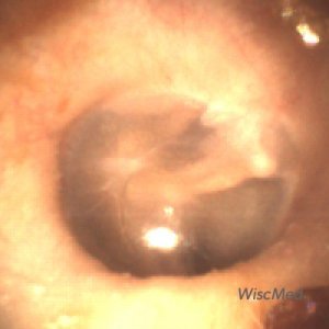
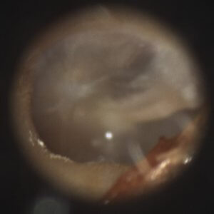
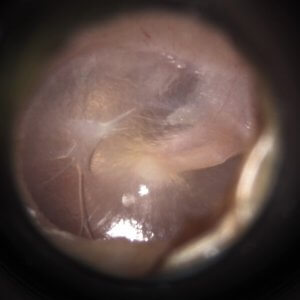
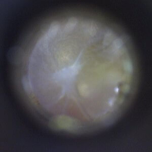
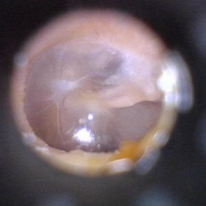
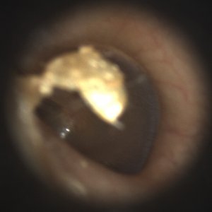
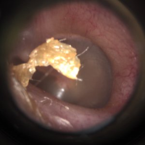
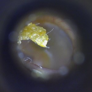
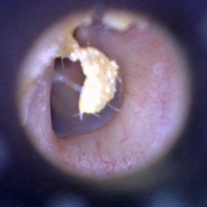
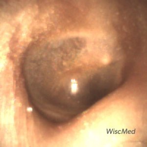
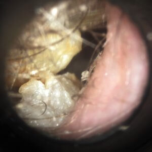
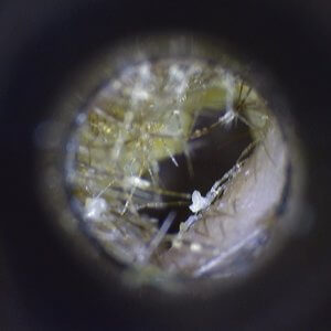
Here are sample videos and images from all five devices in three different scenarios: 1) no ear wax obstruction, 2) mild ear wax obstruction and 3) significant ear wax obstruction. All devices provide a diagnostic image in the case of no ear wax, and in fact higher resolution devices like the Jedmed Horus+, the Welch Allyn digital Macroview and the Teslong are able to provide exquisite detail of the tympanic membrane.
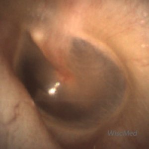
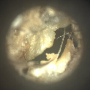
However, as obstruction in the ear canal is increased, with either mild or significant ear wax, the view of the tympanic membrane is either partially or completely blocked for all devices except the WiscMed Wispr. This is the power of the WiscMed Wispr – it’s ability to obtain a diagnostic view of the eardrum in the most challenging ear canals.
No Ear Wax
All Devices provide a diagnostic view of the tympanic membrane. Higher resolution devices provide fine detail. The WiscMed Wispr does not have a tunneling image.
| WiscMed Wispr | Jedmed Horus+ | WA MacroView | Firefly DE500 | Teslong |
 |
 |
 |
 |
 |
Mild Ear Wax
The view of the tympanic membrane is partially compromised for all devices except the WiscMed Wispr.
| WiscMed Wispr | Jedmed Horus+ | WA MacroView | Firefly DE500 | Teslong |
 |
 |
 |
 |
 |
Significant Ear Wax
The WiscMed Wispr is able to navigate through a small window in the wax to provide a diagnostic view of the tympanic membrane.
| WiscMed Wispr | Jedmed Horus+ | WA MacroView | Firefly DE500 | Teslong |
 |
 |
 |
 |
 |
Speculum
In addition to the size and location of the camera, the speculum geometry determines access to the eardrum. The important geometry is both the size of the speculum at the tip, and the presence of a taper. A digital otoscope with a distal camera does not require a taper to the speculum. The speculum is only required for cleanliness between patients. It is not the visual pathway as in traditional analog otoscopy. A speculum without a taper can provide better access to the tympanic membrane. In the photo below you can appreciate that only the WiscMed Wispr speculum has a unique, non-tapered geometry. This allows for the best chance of obtaining a diagnostic image in pediatric ear canals partially obstructed with wax.
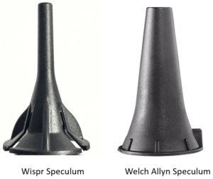
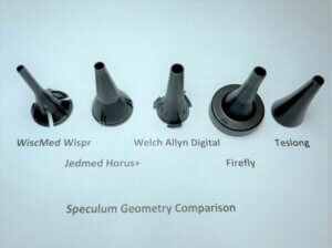
The WiscMed Wispr speculum with it’s unique non-tapered geometry provides the opportunity to navigate through small windows in cerumen.
Clinical Workflow
For an otoscope to be useful in the clinical environment, it must not change the physician’s workflow. The device must be readily available, convenient and with an ease of use like what they are already familiar with. In the clinical environment, the traditional analog otoscope is always close at hand. It is either mounted to the wall, or it is in a charging stand on a desk. Consider where and how you plan to use the digital otoscope. If it is just occasional, “interesting case” use, clinical integration may not be important. It may be acceptable to go to the “special cabinet” to get the device. However, if you want to use the digital otoscope with every patient, integration into the clinical environment will be important to you. A digital otoscope that requires an external computer will be limited to either just one location, or it will need to be on a rolling cart which limits its convenience.
Built-In vs External Display
The advantage of a built-in display is that there is no need to hook up an external device. If the display is not built-in, cables, drivers and a computer or smart phone are required to display the image. This has the disadvantage of being less “handy.” An external computer or display may be satisfactory if you are only using the digital otoscope in one fixed location or only need the digital capability on a limited basis. In addition, a digital otoscope that attaches to an external computer may make integration with the electronic medical record (EMR) easier. A device without a built-in display may not fit into your normal clinical workflow as well. A device with a built-in display will usually be more expensive and potentially may be more fragile, like a smartphone.
Cost
Cost will always be an important factor in choosing a digital otoscope. Consumer digital otoscopes are available from amazon.com for as little as $29. They can provide a high-resolution image of the eardrum. They require external smartphones, applications and cables that reduce their clinical usefulness. In addition, the geometry of the speculum limits access in partially occluded ear canals and they can be uncomfortable. As with most things in life, more features and capabilities result in a higher priced product. Expect to pay more for a built-in display, touch screen, nano camera and speculum. A strong customer support organization, both on-line and via phone also increases product cost.
Comments on Each Device
WiscMed Wispr Digital Otoscope

The WiscMed Wispr Digital Otoscope has the advantage of attaching directly to existing Welch Allyn or Heine handles and containing a built-in touch screen display. This allows it to be conveniently placed wherever an existing analog otoscope is presently located, including on wall mounts and battery handles. The Wispr is unique in the combination of a distal camera and the non-tapered speculum. These features allow for diagnostic views of tympanic membranes in almost all cases except complete obstruction. It is particularly suited to the challenging pediatric population because of the small diameter of the speculum. Images and videos can be reviewed on the device. Pediatric mode builds rapport with the youngest patients. The Wispr provides a diagnostic, but not a high-resolution image of the eardrum. Because of the location of the camera at the end of the speculum, it is susceptible to debris and must be wiped down with an alcohol pad between each patient.
Pros; Attaches to existing handles, built-in touch screen, no-taper speculum with distal camera prevents tunnel image, able to obtain diagnostic images of the tympanic membrane except in cases of complete obstruction. Pediatric Mode. Dedicated support organization.
Cons; Distal camera susceptible to debris, diagnostic but not high-resolution image.
If you would like to know more about the construction of the WiscMed Wispr digital otoscope, please see our $995 value on Wispr University.
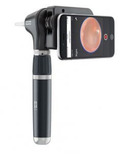
This is an analog otoscope that has an attachment for your smartphone camera via an interface plate. The interface plate connects the analog otoscope and your smartphone using double-sided sticky tape. This attachment method is insecure and does not allow for removing of the phone between uses. Since every smartphone has a different camera arrangement, the interface plate must be quite large to allow for alignment of the proper camera on the smartphone with the interface plate aperture. The device is heavy, with a smartphone hanging awkwardly near the handle. It is difficult to be able to press the capture button on the application while stabilizing the ear exam. The application on the smartphone is complex and difficult to use while performing an exam. The speculum is large, without taper and the camera on the smartphone is distal to the tip of the speculum. The result is a tunnel image with poor ability to navigate past obstructions.
Pros; From well-known medical device company. Standard speculum.
Cons; Hard to setup. Poor attachment of phone using tape. Attachment is not clinically robust. Bulky and heavy with poor ergonomics. Tunnel image. No ability to navigate past cerumen. Difficult to optimize lighting.
JedMed Horus+ HD Video Otoscope

The Jedmed Horus+ HD Video Otoscope is the only standalone device reviewed here. It has a built-in display and battery. In addition to the built-in display, it is also benefits from being able to attach to an external display. It provides a high-resolution image, and the manual focus is straight-forward. Images and videos can be reviewed on the device. Because of the proximal camera and the speculum taper, the image will suffer from tunneling, and any amount of wax will prevent a good view of the eardrum. Although the device has a touch panel below the display, the display itself is not a touch screen. Changing functions on the device such as image capture to video capture requires navigating a menu system via the touch panel.
Pros; Built-in display, self-contained with battery power, high resolution, able to attach to external display.
Cons; Proximal camera, tapered speculum, tunnel image, moderate wax will block visualization of the eardrum, touch panel but not touch display.
Welch Allyn Digital MacroView Otoscope

The Welch Allyn digital MacroView otoscope brings digital capability to a familiar form factor by a leading provider of medical equipment. It delivers high resolution images using standard speculum that leads to a tunneling effect. There is no video lag and focus is logical. The MacroView requires both a Welch Allyn handle and a connection to a computer. The requirement of external cables and a computer limit its clinical usefulness to positioning in a single room. The requirement of the computer will also affect normal clinical workflow. Because of the proximal camera and the speculum taper, the image will suffer from tunneling, and any amount of wax will prevent a good view of the eardrum.
Pros; leading medical device provider, high resolution, familiar form factor, “standard” speculum
Cons; cost, requires both a Welch Allyn handle and external computer, tunnel image, moderate wax will block visualization of the eardrum

This is the least expensive digital otoscope that attaches to an external computer as a display. It can provide a high-resolution image. It is manufactured by a company that makes a broad line of imaging products. It is shaped like a fat cigar, and the DE500 suffers from a notable video lag which when combined with its manual focus makes it difficult to use. Any amount of wax will prevent a good view of the eardrum.
Pros; least expensive solution with external computer, high resolution
Cons; external cables and computer, proximal camera, large speculum size and taper, tunnel image, significant video lag, poor ergonomics, moderate wax will block visualization of the eardrum
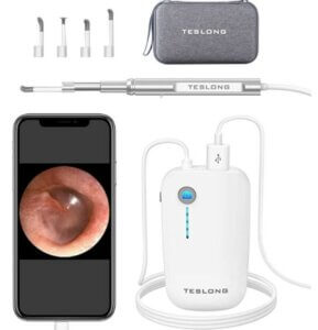
This is a good fit for casual home use or if you want an inexpensive otoscope and do not mind having cables and an eternal smart phone for display. It provides a high-resolution image of the eardrum, but only in cases of no wax. Any amount of wax will preclude a good view of the eardrum. It may have some application in teaching environments but will likely not provide a reliable image in most pediatric ears. It may not be as comfortable for the patient as other digital otoscopes. It will not be durable in a clinical environment, and It is not a good fit for a busy clinic.
Pros; low cost, high resolution
Cons; external cables and smartphone, poor ergonomics, speculum size and taper, tunnel image, moderate wax will block visualization of the eardrum
James Berbee, M.D.
Emergency Physician
Founder, WiscMed
June 2021
jim.berbee@wiscmed.com
www.wiscmed.com

