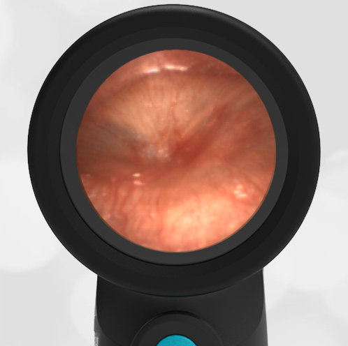
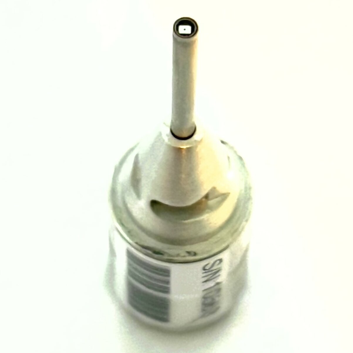
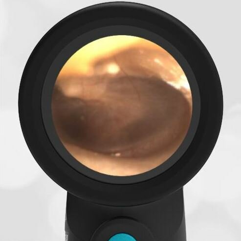
fbpx


Wispr University

Welcome to Wispr University! Here you can expect to find unique insights and educational content about the ear exam including anatomy, pathologies, treatment and techniques as well as an insider look at the Wispr Digital Otoscope and the unique ways that the Wispr can be utilized in your practice. At Wispr University, our goal is to be a place of learning that helps to advance the ear exam in the practice of medicine. Be sure to stop by often, we will be providing frequent (and interesting) updates. We’ll see you around campus!

chorda tympani image

Normal Middle Ear Anatomy as Seen by Otoscopy

January 3, 2022
The human ear is an example of nature’s amazing bioengineering. It translates sound waves first into mechanical motion and then into electrical signals that are interpreted as sound by the brain. The otoscope can “see” the tympanic membrane (eardrum) which separates the external ear from the middle ear. Because the eardrum is...Read more about Normal Middle Ear Anatomy as Seen by Otoscopy
Day-0-Label-left-aspect-ratio-1-1

Progression to Acute Otitis Media (AOM)

December 28, 2021
Read more about Progression to Acute Otitis Media (AOM)
Jim Berbee Clinic Pic (3)

Acute Otitis Media (AOM) – a spectrum of images from normal to AOM

June 25, 2021
Read more about Acute Otitis Media (AOM) – a spectrum of images from normal to AOM
Wispr Digital Otoscope by WiscMed showing ear exam image of Acute Otitis Media AOM by Dr. Daniel Ostrovsky

Seeing is Believing Part 2 – Acute Otitis Media

March 1, 2021
Read more about Seeing is Believing Part 2 – Acute Otitis Media
Dr. Gregory S. Rebella WiscMed Co-Founder

Seeing is Believing Part 1 – Challenges to Improving Diagnostic Accuracy

January 25, 2021
Challenges to Improving Diagnostic Accuracy – Part 1 Read more about Seeing is Believing Part 1 – Challenges to Improving Diagnostic Accuracy
OMA-w-camera-focus-1-aspect-ratio-1×1

Digital Camera Location

December 30, 2020
There are a number of choices available for digital otoscopes. We have previously reviewed 5 digital otoscopes here . While the basic components of each of these devices are the same – a digital camera, a light source and a display – how these components are implemented makes a vast difference in the clinical effectiveness…Read more about Digital Camera Location
12032020-left-aspect-ratio-1×1

More than Acute Otitis Media!

December 5, 2020
Read more about More than Acute Otitis Media!
Wispr-in-Ear-Canal-aspect-ratio-1×1

Wispr Digital Otoscope Exam Tips

December 2, 2020
This video provides tips for a successful ear exam using the Wispr digital otoscope How to perform a digital otoscope exam Make sure the camera is clean. Test the camera by pointing it at text on paper. The text should be sharp and readable. Make sure the speculum is firmly attached. The speculum should not…Read more about Wispr Digital Otoscope Exam Tips
09302020-II-Pop-aspect-ratio-1×1

Valsalva Maneuver – Ear Popping

November 22, 2020
Read more about Valsalva Maneuver – Ear Popping
Pneumatic-Otoscopy-aspect-ratio-1×1

Pneumatic Otoscopy

November 17, 2020
Read more about Pneumatic Otoscopy