Wax Annulus – May 4, 2023
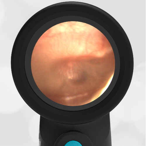
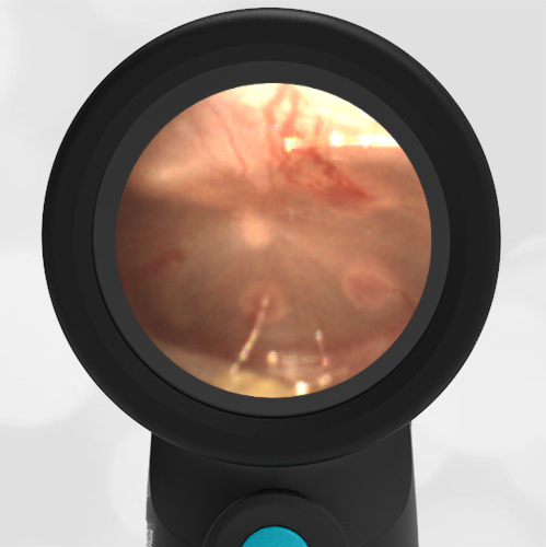
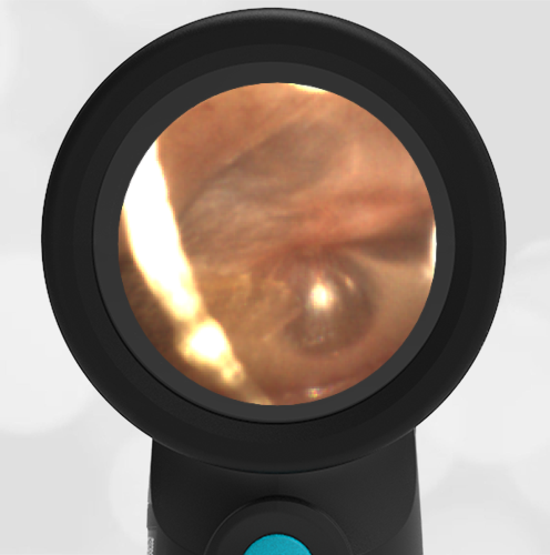
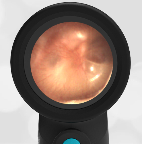
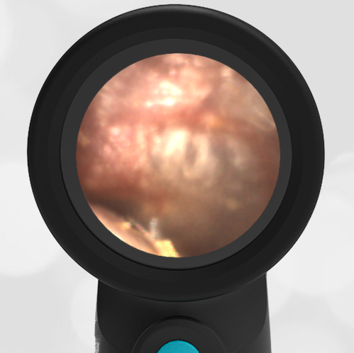
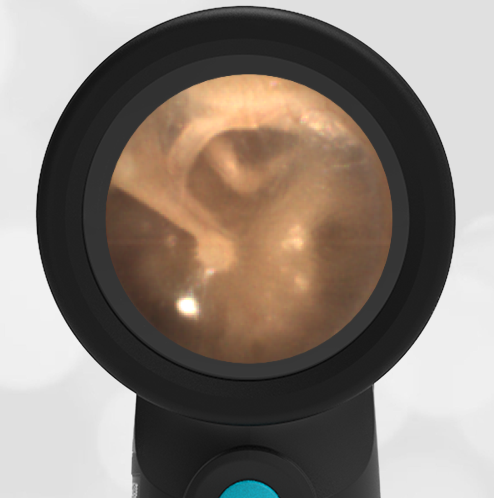
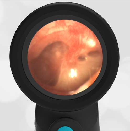
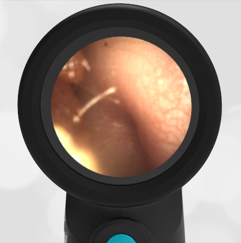
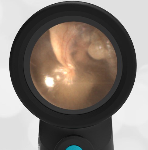
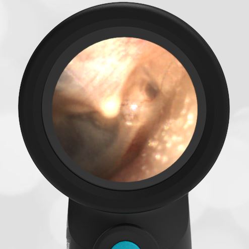
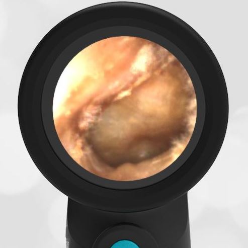
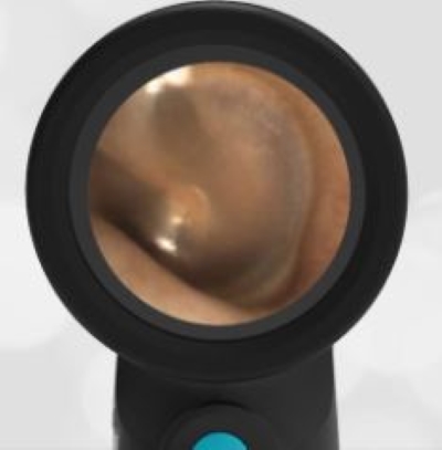
A healthy 40-year-old male presents to internal medicine for a routine physical exam. He has no concerns about today’s visit. The following image of his left ear is obtained with the Wispr digital otoscope. What is your diagnosis, and what is the next step in management?
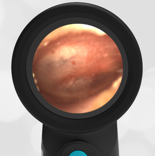
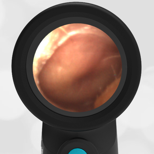
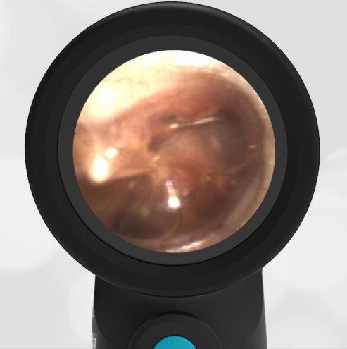
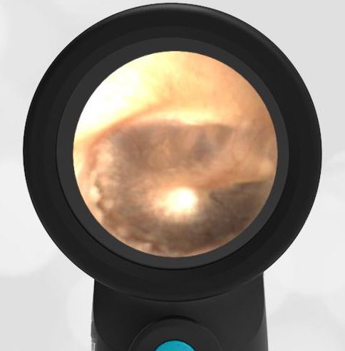
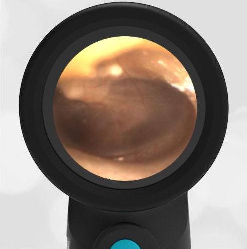
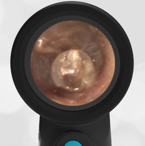
The patient has a ring of cerumen (wax) lying inferiorly. No further action is necessary.

This is an interesting image. At first glance, it appears to be a tympanic membrane rupture. However, it is simply cerumen (wax) layering inferiorly. The cerumen in this image has a lighter and almost transparent appearance that makes it look like it could be the tympanic membrane, and thus a rupture. Actually, the cerumen is lying inferiorly from about the 10 o’clock to 3 o’clock position. There also appears to be a partial ring of cerumen that is deeper at the 11 o’clock to 3 o’clock position.
This digital image allows time to realize that the expected features of the tympanic membrane are present and undisturbed. These features include the malleus and incus, the cone of light, and the Eustachian tube (ET) shadow. This is an uncommon appearance of cerumen. Generally, ear wax appears more yellow.
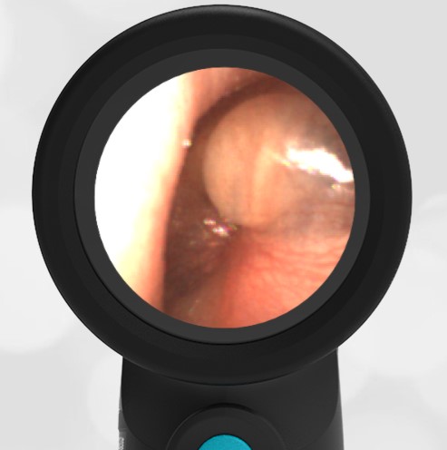
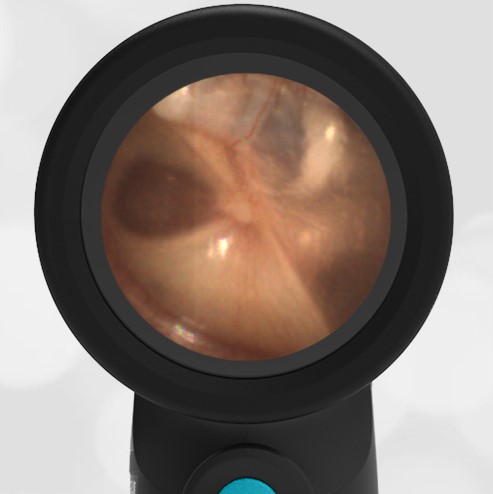
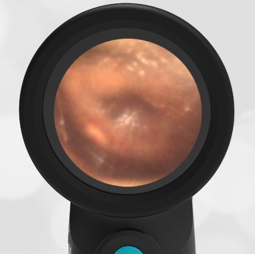
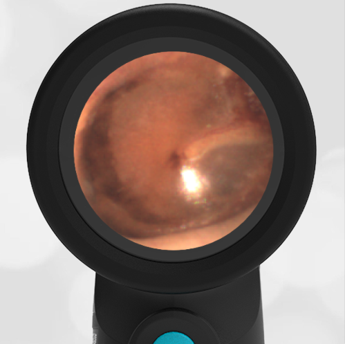
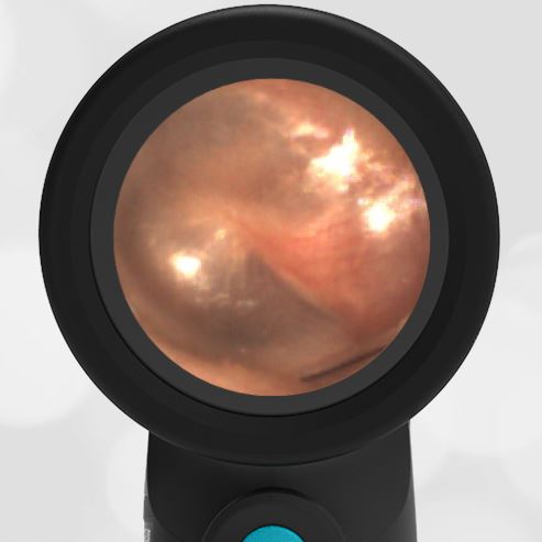
Here is the video of the complete ear exam: