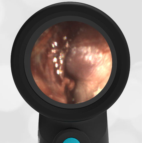
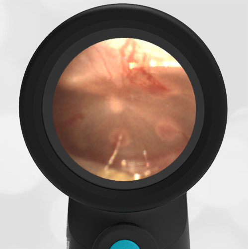
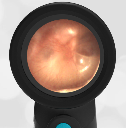
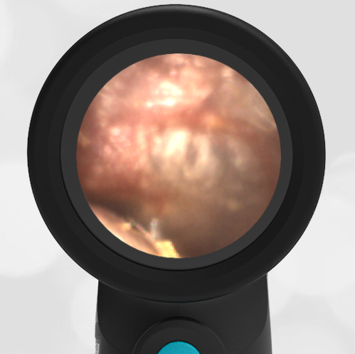
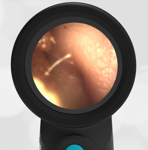
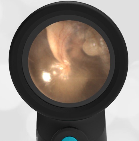
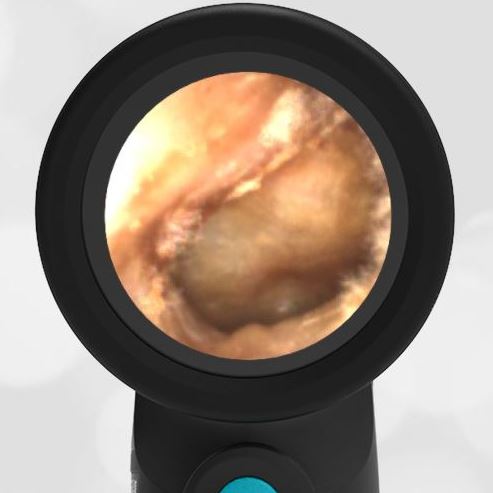
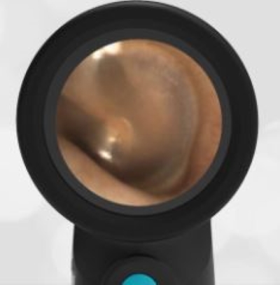
Extruded and Encrusted Ventilation Tube
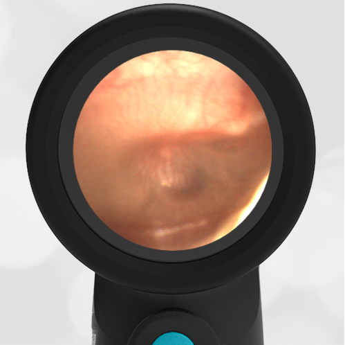
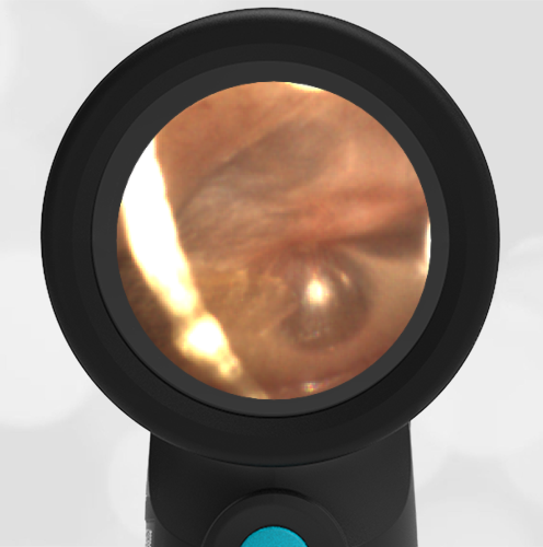
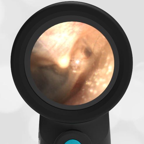
An otolaryngology (ENT) resident stopped by the WiscMed booth at the 2021 ENT OTO Experience conference in Los Angeles, CA to experience the Wispr Digital Otoscope. The Wispr Digital Otoscope was used to perform an exam on her right ear. This image was obtained. What is your diagnosis?
The resident has an extruded and encrusted ventilation tube.
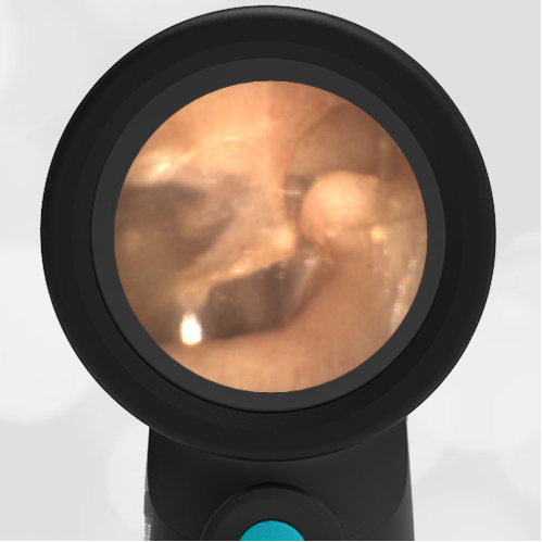
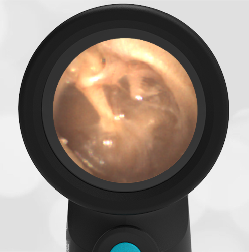
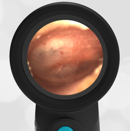
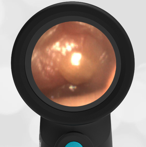
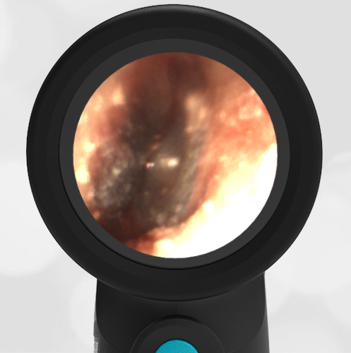
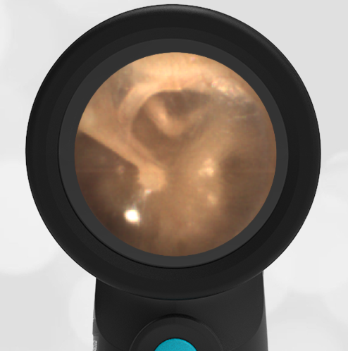
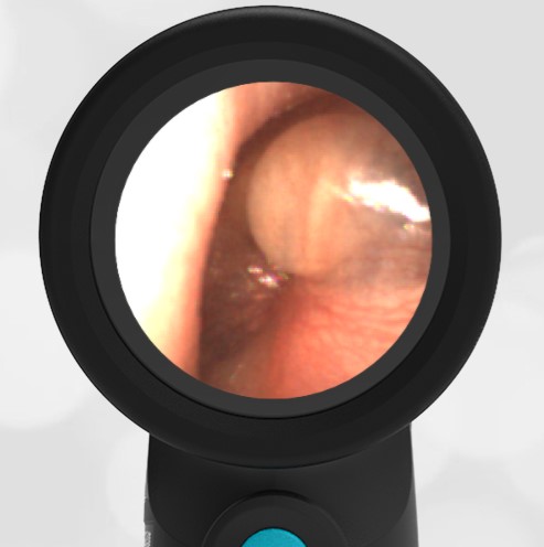
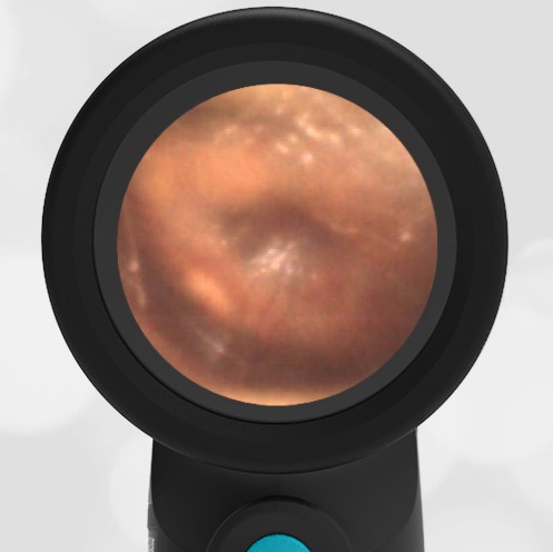
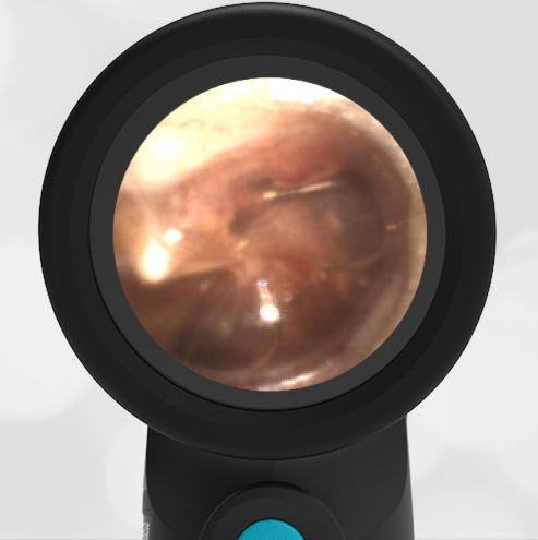
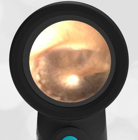
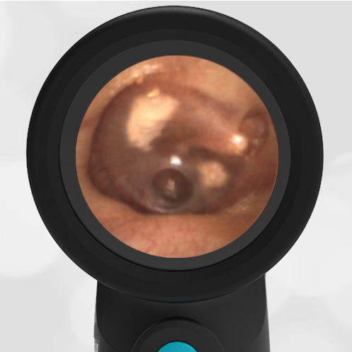
After obtaining this image and further history, the ENT resident endorsed having a tympanostomy (ventilation) tube placed in her right ear about a year ago for what was described as an effusion. Generally, an effusion would not be an indication for ventilation tube placement, but further information regarding the diagnosis resulting in the tube placement was unavailable. Tympanostomy tubes are generally placed in cases of repeated otitis media with eustachian tube dysfunction. The tubes are placed in the anterior inferior quadrant of the pars tensa, which lines up nicely with the Eustachian tube in the middle ear. They generally “fall out” on their own in about a year. Here is another example of an extruded ventilation tube.
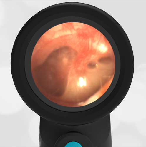
Take a look at the complete video of this exam which demonstrates the power of the Wispr Digital Otoscope to help with diagnosis. Note how the distal camera on the Wispr is able to obtain a clear view of the annulus of the ventilation tube, confirming the object. The ventilation tube is most likely encrusted with keratin and cerumen (ear wax) which is why the nature of the object was initially unclear.
Complete exam video