Acute Otitis Media (AOM)
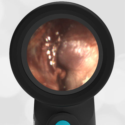
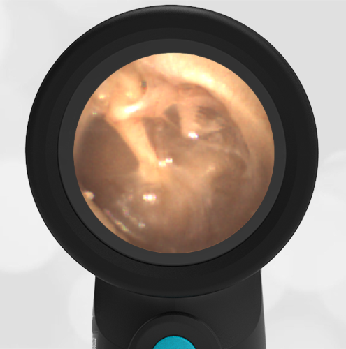
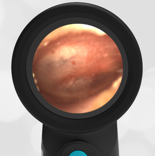
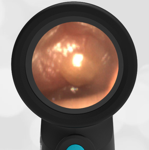
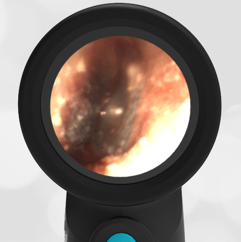
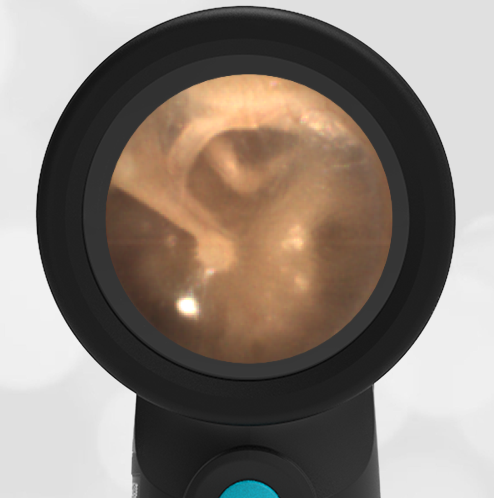
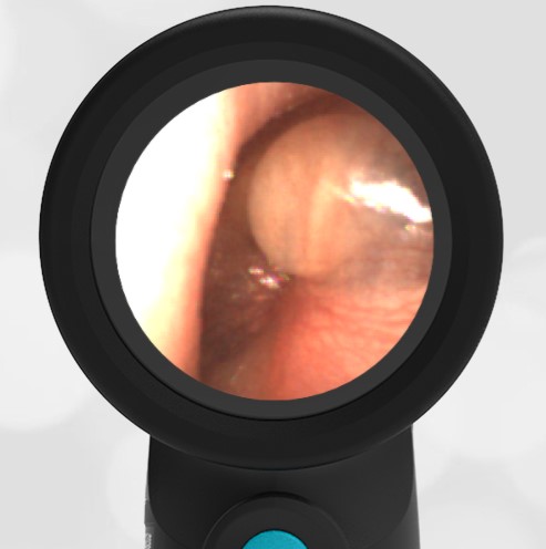
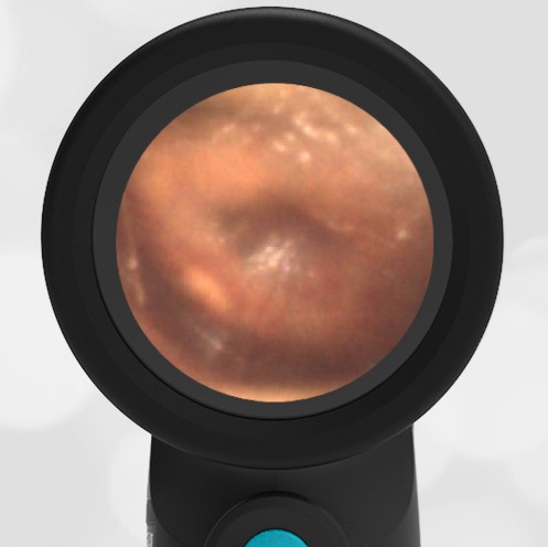
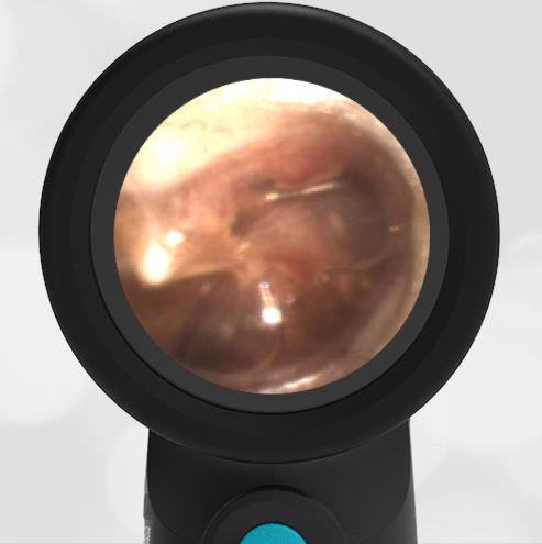
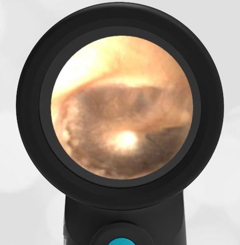
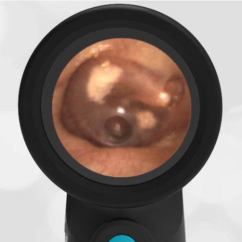
A 4-year-old female with a history of ear infections presents to the pediatric clinic with 3 days of rhinorrhea, malaise, and temperature to 102.3 F. Several hours ago, the child began to complain of right ear pain. The mother has been treating the child with Tylenol. The following image is obtained from the right ear.
The child has acute otitis media (AOM).
You can also see a clear air-fluid level behind the tympanic membrane in the superior-anterior (right-upper) location. The key diagnostic feature of AOM is a bulging tympanic membrane as seen in this image. As also seen in this image, there is often a loss of the malleus bony landmark and of the cone of light. Erythema (redness) is usually present. This may be an early presentation of AOM as the malleus bone is still partially recognizable. Compare this presentation to otitis media with effusion (OME). Both have air-fluid levels and some erythema. The difference between AOM and OME is the bulging of the tympanic membrane. AOM is usually treated with antibiotics while OME is observed. The air-fluid level is caused by fluid behind the tympanic membrane that isn’t draining properly due to inflammation of the eustachian tube. If the child has significant recurrent infections, tympanostomy tubes may be considered.
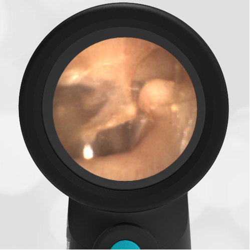
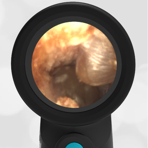
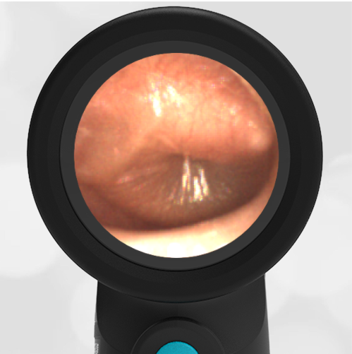
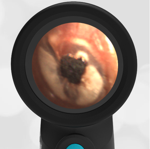
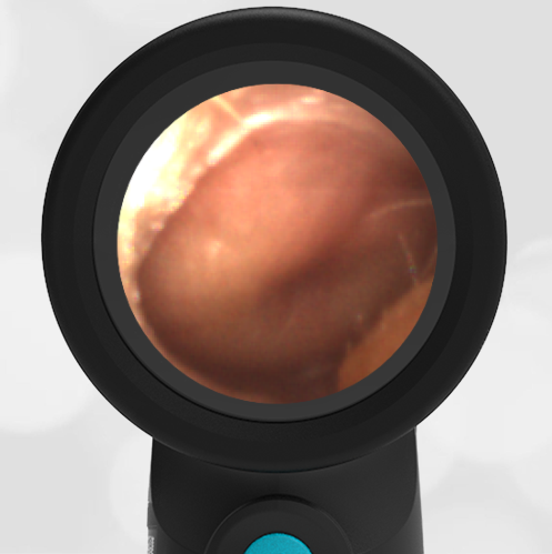
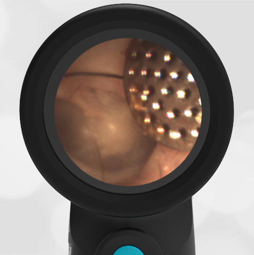
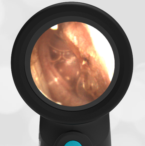
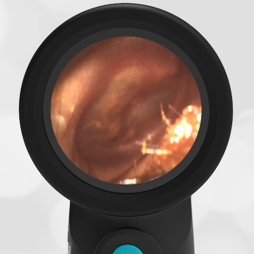
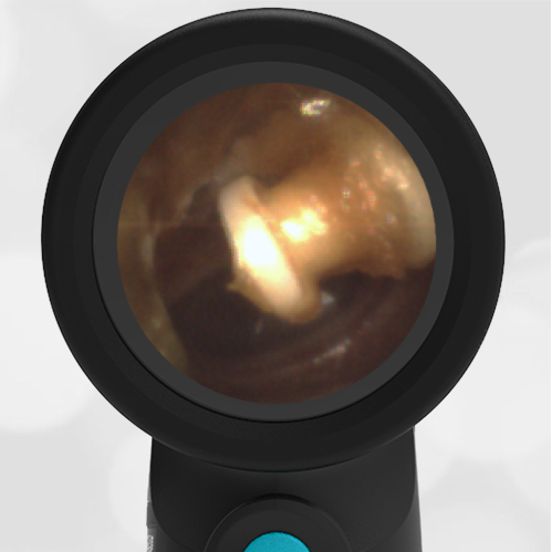
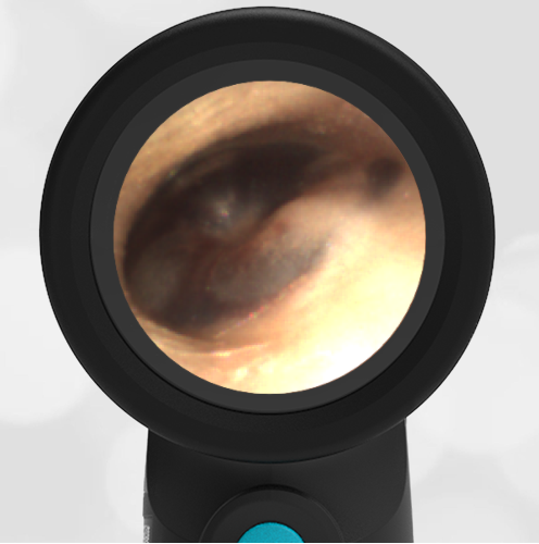
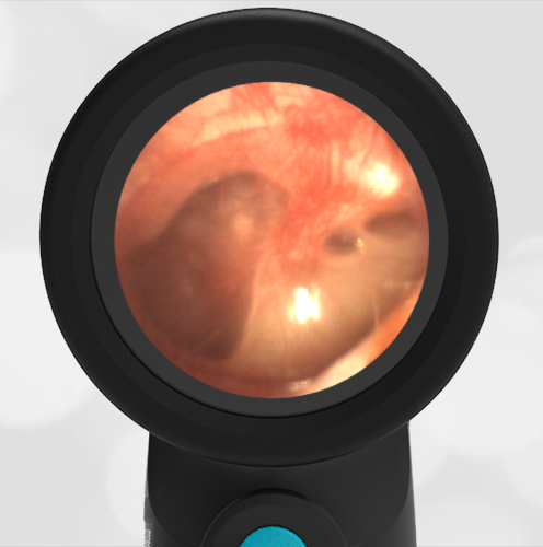
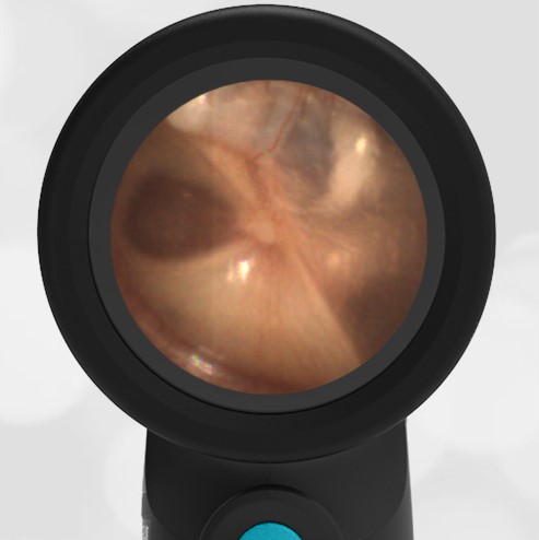
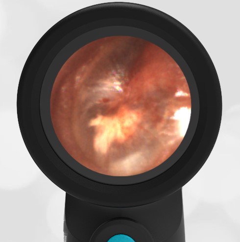
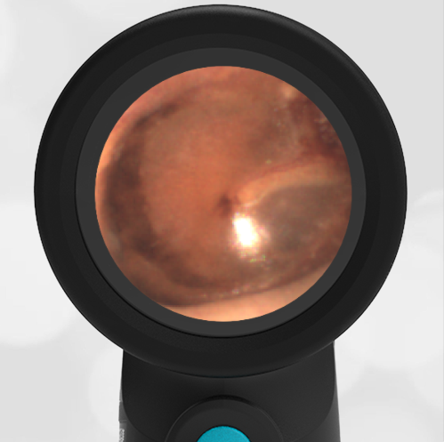
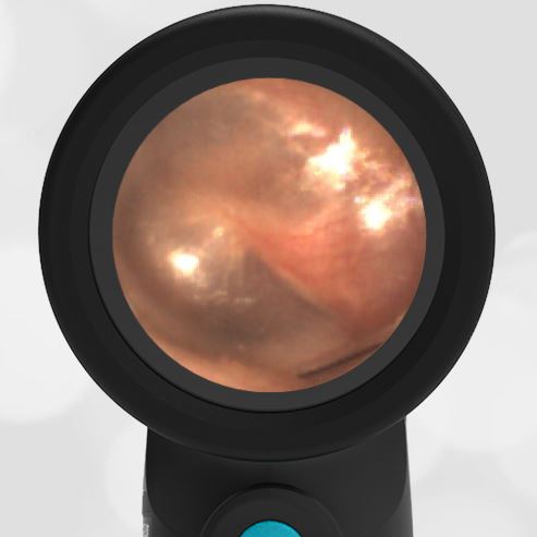
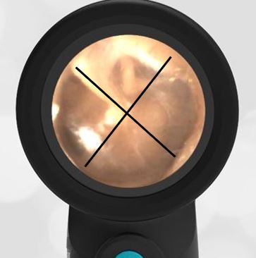
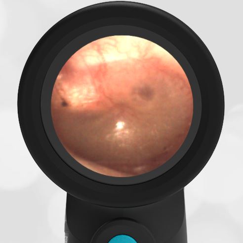
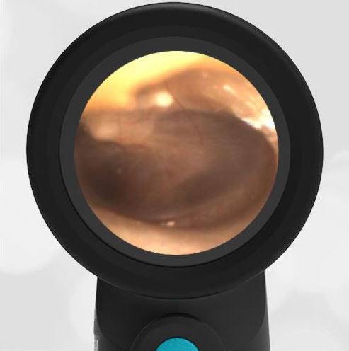
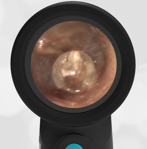
Here are two additional examples of acute otitis media.
-
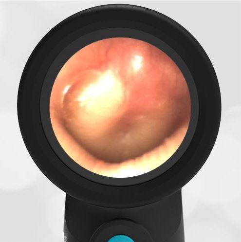
- Severe Bulging
-
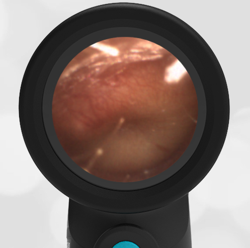
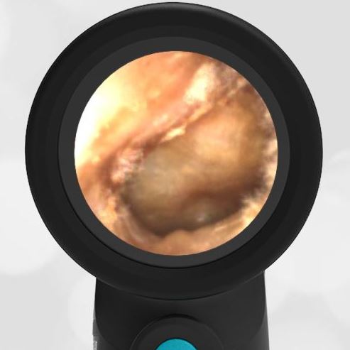
- Acute Otitis Media (AOM)
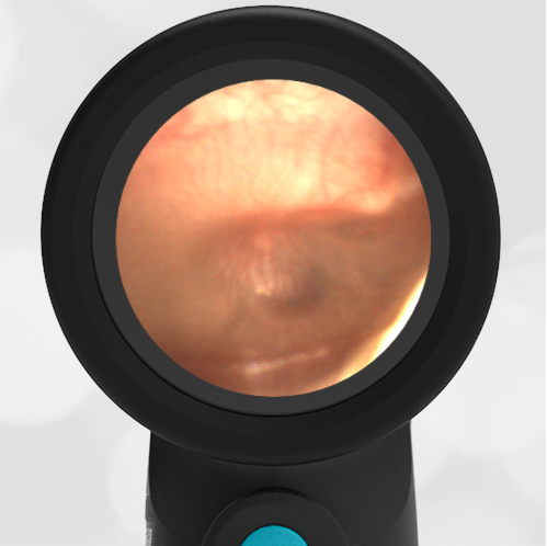
A presentation on normal ear anatomy is available at Wispr University.