fbpx


Cases

Middle Ear Effusion

Middle Ear Effusion

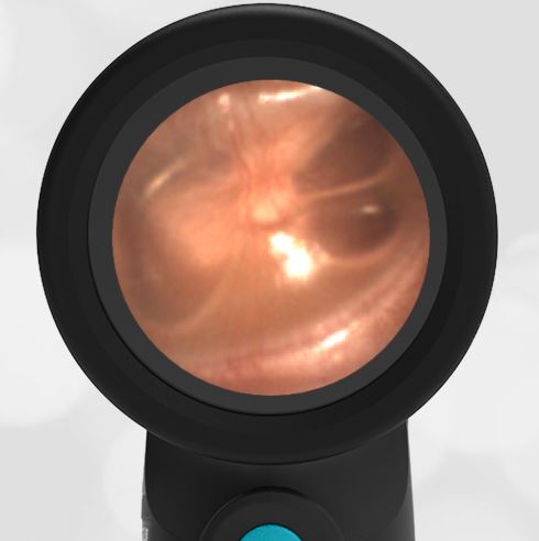
A 12-year-old female presents to the pediatric clinic for a routine well-child appointment. The patient reports that a few days ago she had a cough, runny nose, and congestion. Today, she says the symptoms have resolved and she feels well.


This image of the right ear was obtained.

What is true about this condition?

  1. It is caused by a blockage in the Eustachian tube.
  2. The source is bacterial.
  3. Tympanostomy (ear) tubes are indicated.
  4. No intervention is needed.

The answer is 4. No intervention is needed.

The child has a middle ear effusion.

This is a common clinical scenario, an incidental finding on a physical exam. The ear is not normal in that there is fluid behind the tympanic membrane (eardrum). This is referred to as a middle ear effusion.

The presence of air-fluid levels suggests both inflammation of the middle ear space and proper ventilation. The inflammation is no doubt due to sequala of the minor viral syndrome the patient has described recovering from. The air-fluid levels let us know that the middle ear space is being properly ventilated by the Eustachian tube. There is a mild bulging of the pars flaccida portion of the TM. This is the ”loose” portion of the TM and responds to even a small increase in middle ear pressure. The important finding is that the TM is not bulging, and the malleus bony landmark is clearly identifiable.

Contrast the middle ear effusion with acute otitis media (AOM) below. Not the marked bulging across the entire TM in the case of AOM. There are also no air-fluid levels in AOM which means the middle ear space pressure is not being equalized via the Eustachian tube.

Middle ear effusions typically resolve in days to weeks. No treatment is generally needed. Here are two other examples of middle ear effusions – one, two.