Fibrous disk
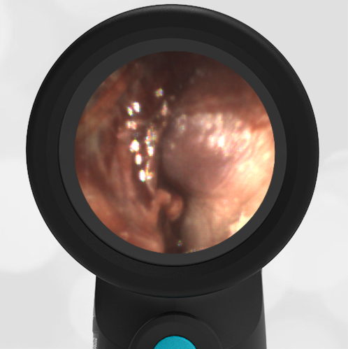
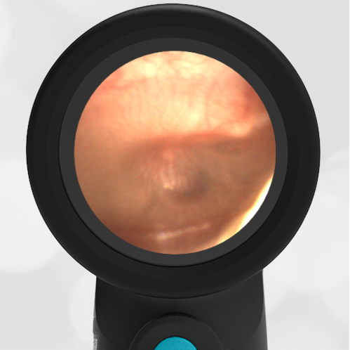
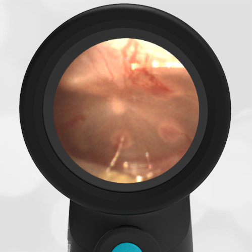
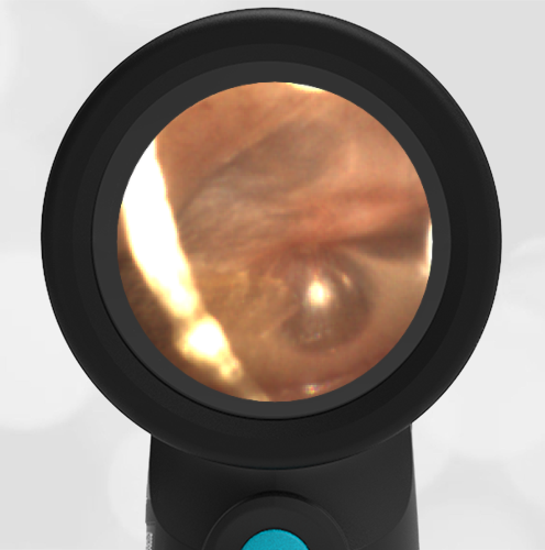
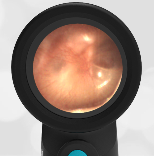
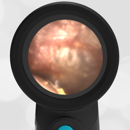
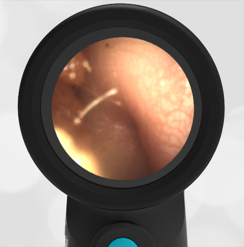
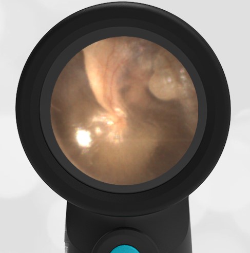
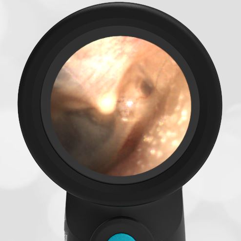
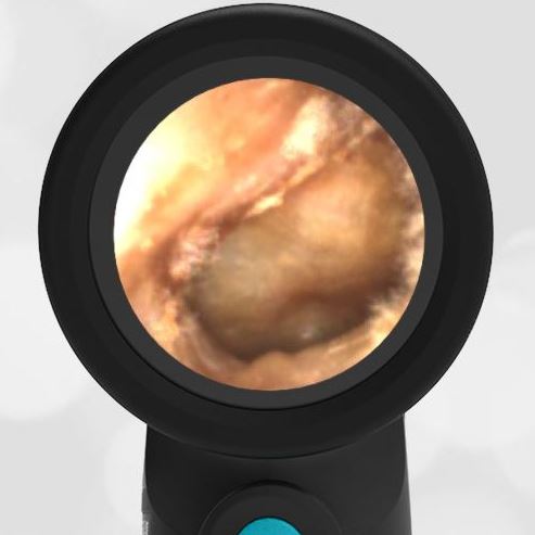
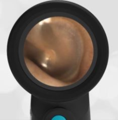
A 42-year-old male is seen by his primary physician for a routine checkup. He has no complaints. This image of his right ear is obtained.
What abnormalities do you appreciate and what interventions are indicated?
The patient has a fibrous disk along with pseudo retraction of the pars flaccida. No intervention is indicated.

This image shows the middle ear anatomy nicely. The malleus and incus ossicles are clearly visible. The chorda tympani nerve can also be identified although it is not labeled due to space constraints. This image has several features that distinguish it from a “normal” tympanic membrane. The most obvious is the large fibrous disk. The tympanic membrane (eardrum) is comprised of three layers of tissue. The (1) outer cutaneous layer, the (2) middle fibrous layer, and the (3) inner mucosal layer. In the case of this patient, it appears that there is a localized thickening of either the outer cutaneous or middle fibrous layers. The cause of this is not known, but it does not seem to involve either infection or trauma. It is likely of embryologic origin.
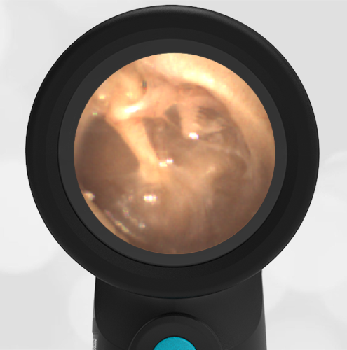
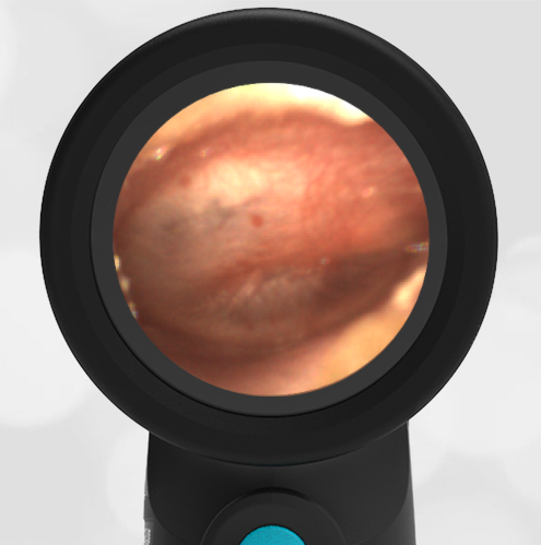
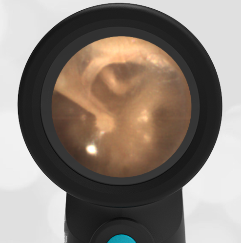
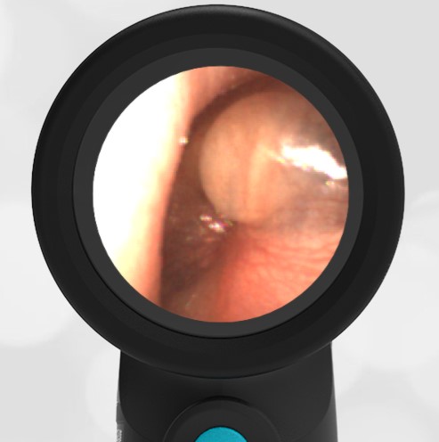
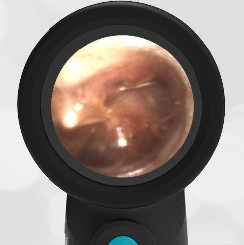
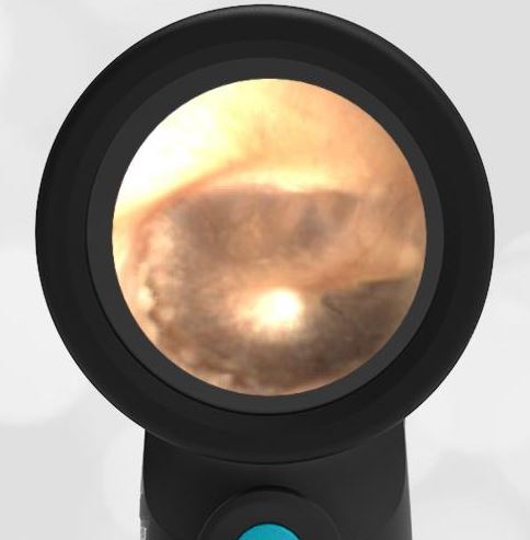
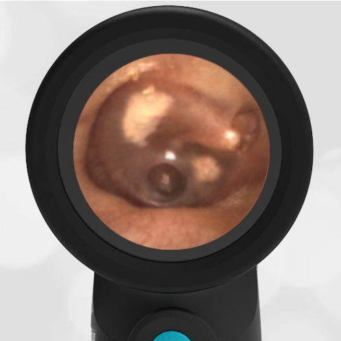
In addition to the fibrous disk, there is also pseudo retraction of the pars flaccida portion of the tympanic membrane. The tympanic membrane divides the external ear from the middle ear. The portion of the membrane that drapes across the superior portion of the malleus bone can become retracted for several reasons. One reason is a prior history of trauma or infections, the other is compressed geometry of the ear canal. In this case, the compressed geometry of the ear canal is likely causing a fold of the tympanic membrane. This might be considered a pseudo retraction. Compare with a pars flaccida retraction.
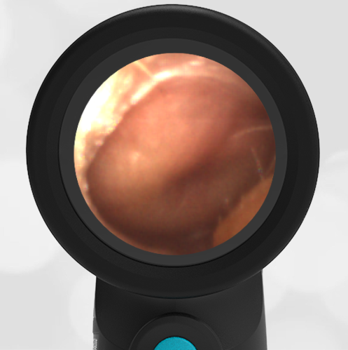
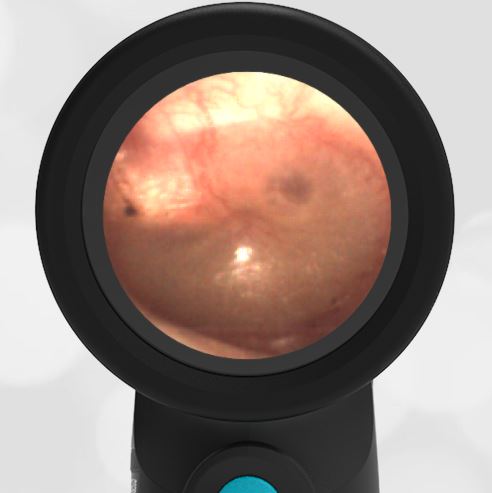
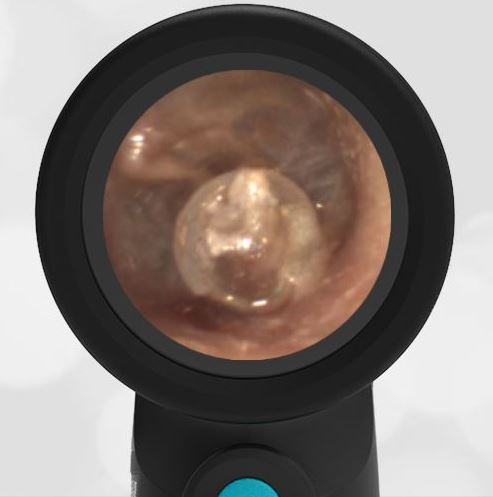
Neither the fibrous disk nor the pseudo pars flaccida retraction are concerning. They likely have no effect on hearing. No intervention is needed.