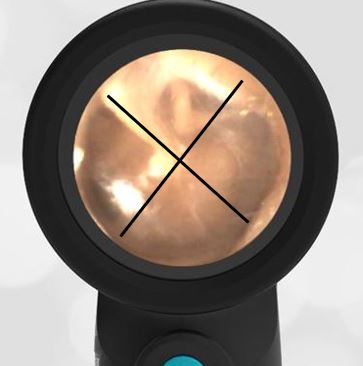
What is Your Advice?
A medical student is rotating in the pediatric clinic. She shows her attending the following three ear images. She wants to start antibiotics on all three patients.
Do you agree that antibiotics should be started?
Besides its ability to obtain reliably diagnostic images of the tympanic membrane, the Wispr digital otoscope is also good for sharing images with learners, patients, and families. These three images demonstrate commonly observed otoscopic findings.
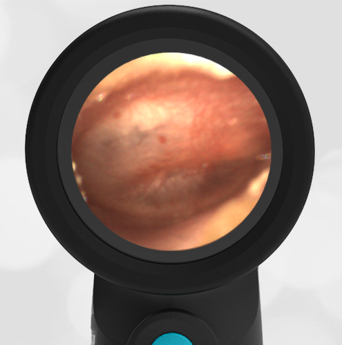
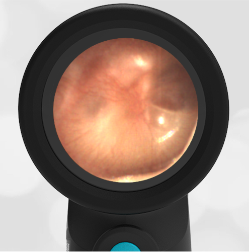
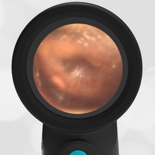
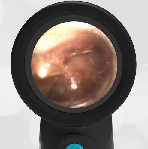
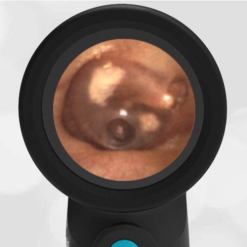
Myringosclerosis
Sclerosis
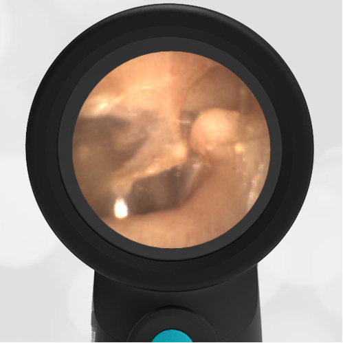
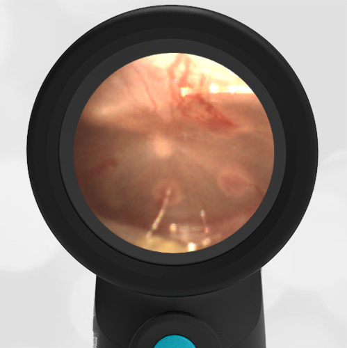
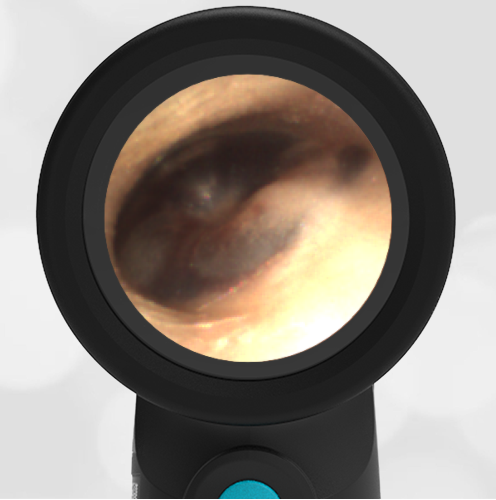
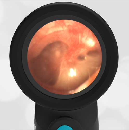
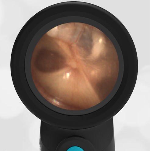
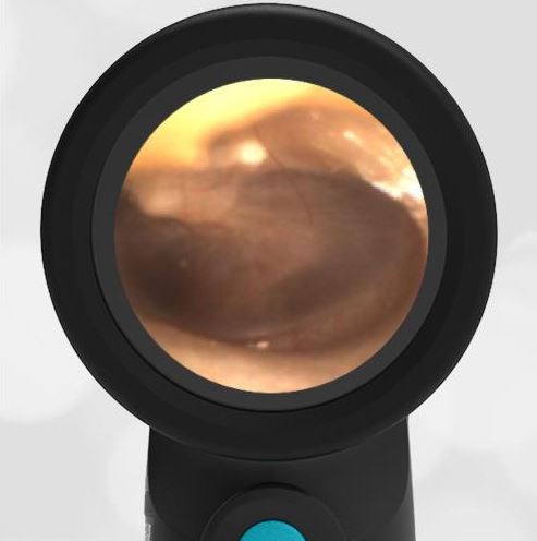
The first image is of myringosclerosis (sclerosis). This is localized thickening (calcification) of the eardrum, generally observed to be white, as in this case. The sclerosis is generally from an eardrum that has been damaged and then healed. Common causes of damage include tympanostomy (ventilation, ear) tubes and traumatic rupture of the eardrum. Sclerosis, although often dramatic, does not generally materially affect hearing. Sclerosis is not an indication to start antibiotics. It represents a well-healed eardrum and no further intervention is necessary.
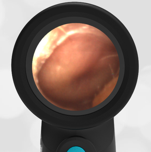
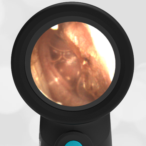
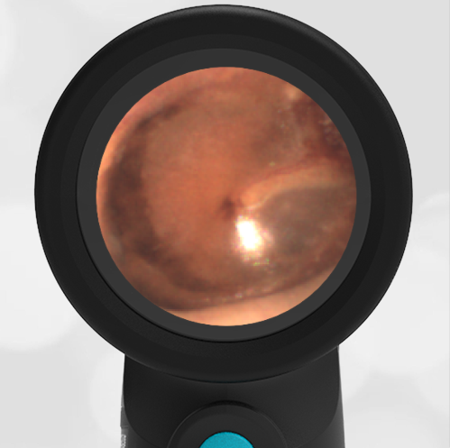
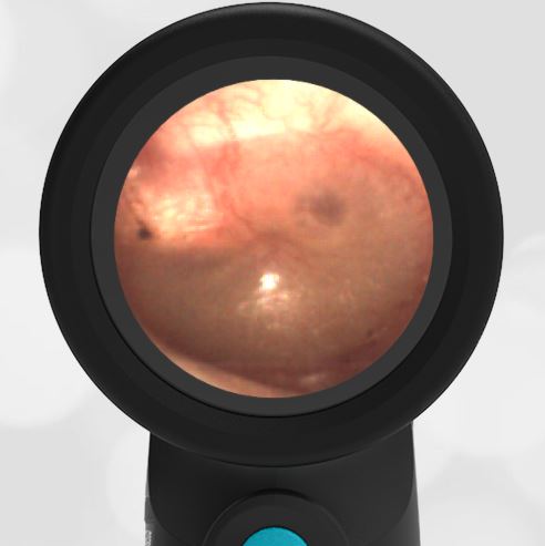
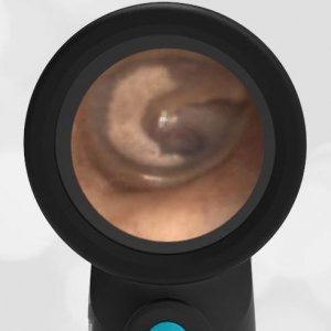
Tympanostomy (Ventilation, Ear) Tubes
Ventilation Tubes
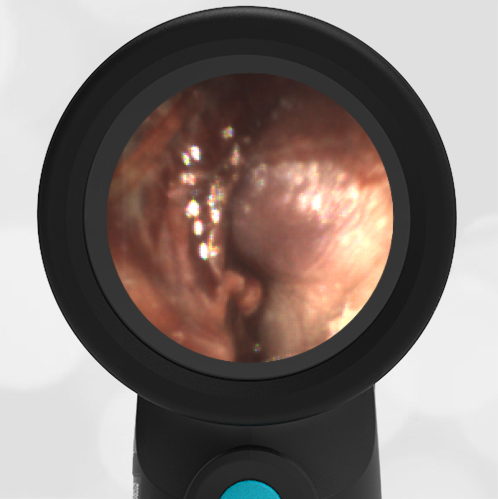
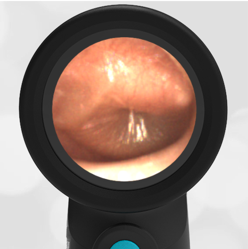
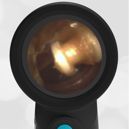
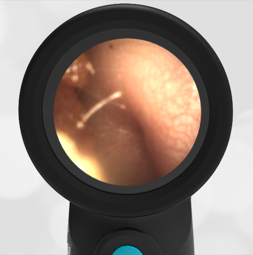
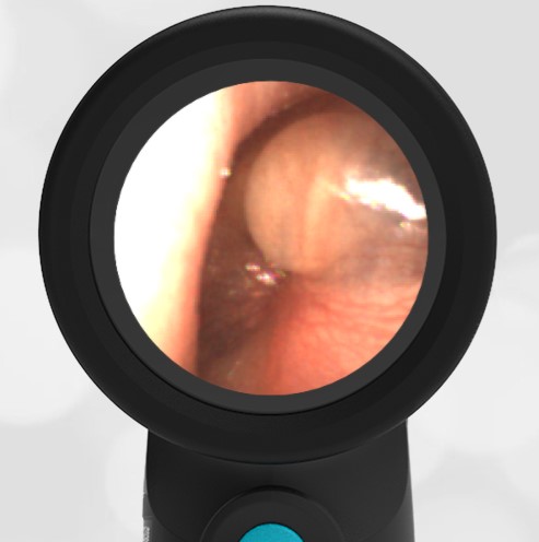
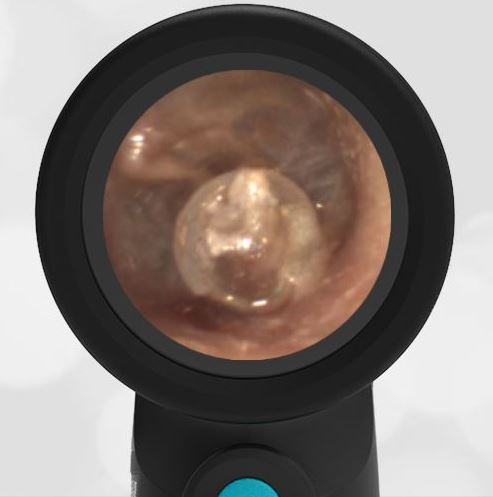
The second image shows a well-positioned tympanostomy tube (blue circle) in the anterior-inferior portion of the pars tensa tympanic membrane. There is a minor amount of debris around the annulus of the tube that might be cerumen (ear wax) or it could be drained and congealed middle-ear fluid. The rest of the eardrum is completely normal with a pearly grey light reflex, concave surface and the malleus bone clearly visible.
Ventilation tubes are inserted to drain the middle ear space. They are generally placed in children who have recurrent acute otitis media (AOM). The tubes fall out on their own in months to years. After the ear tube falls out, there is generally an area of sclerosis present as above. A ventilation tube in an otherwise normal-appearing ear is not an indication to start antibiotics.
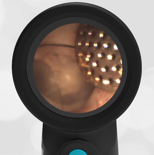
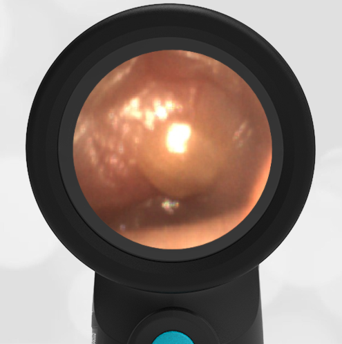
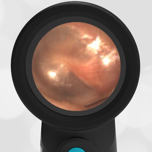
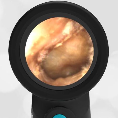
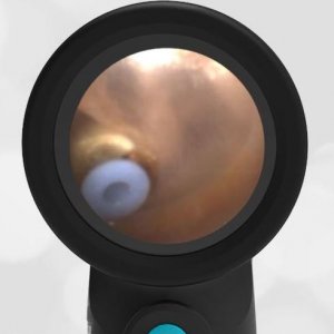
Acute Otitis Media (AOM)
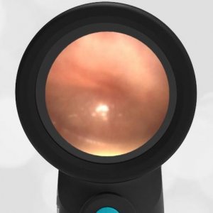
Acute Otitis Media
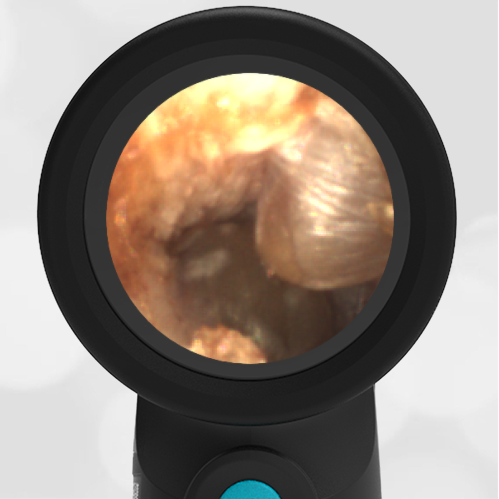
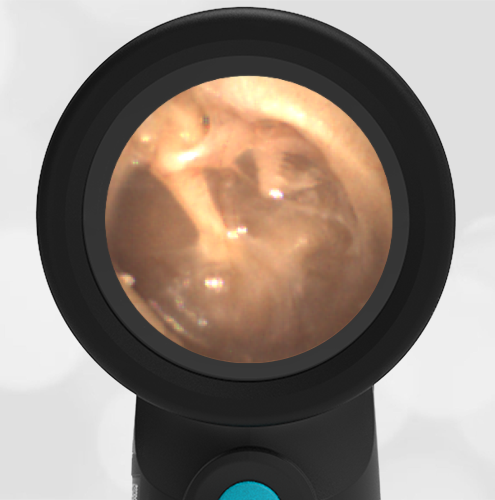
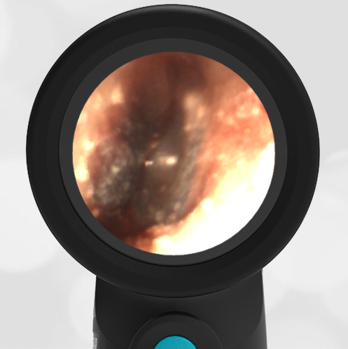
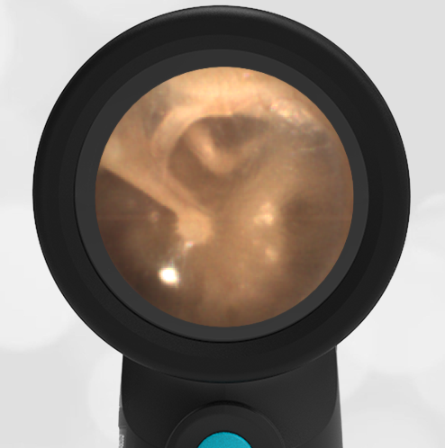
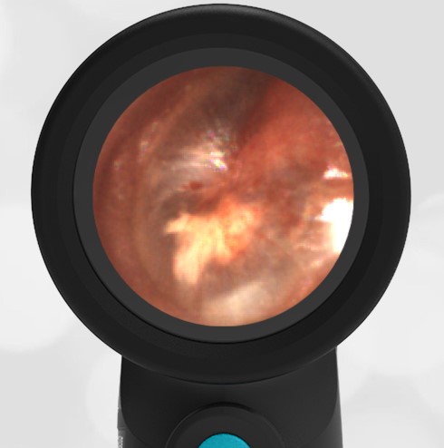
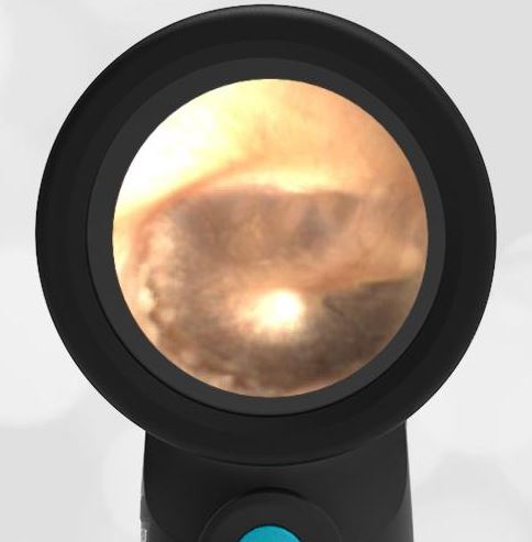
The third image shows a mild case of acute otitis media. There is bulging of the pars flaccida and the superior-posterior portion of the pars tensa tympanic membrane. There is a partial loss of the malleus bony landmark. This case appears to be mild because the bulging is not severe in the inferior portion of the tympanic membrane. The decision to start antibiotics, in this case, is indeterminate. It would depend on how ill the child is and how long he has had the symptoms. Of the three images presented, this is the only case where antibiotics should be considered.