Acute Otitis Media (AOM)
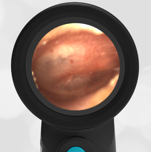
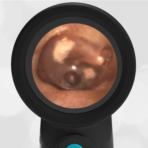
A 3-year-old female presents to urgent care with her parents for evaluation of fever and ear pain. The parents report that the child has had three days of viral symptoms including cough, runny nose, and a temperature to 38 C. They have been treating her with over-the-counter analgesics to which she has responded well. This morning the child began to complain of ear pain. She does not have any history of ear infections and has been well. The following image is obtained with the Wispr digital otoscope.
What is your diagnosis and treatment?
The child has acute otitis media (AOM) and antibiotics should be considered.
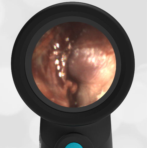
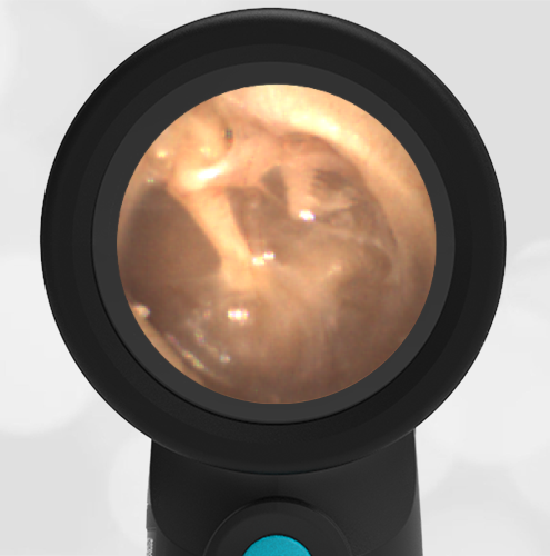
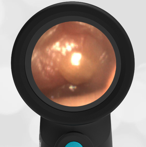
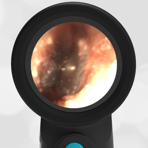
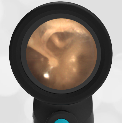
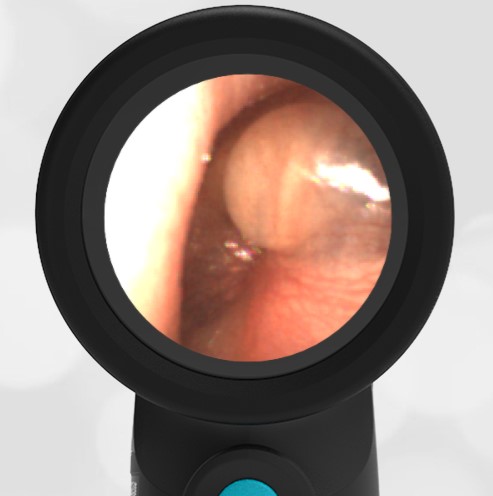
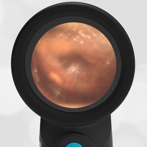
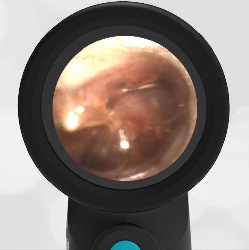
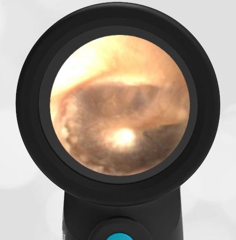
This is a very common presentation in acute care. A pediatric patient with a prodrome of viral symptoms advancing to ear pain. The Wispr digital otoscope image shows all the features of acute otitis media (AOM). There is bulging of the ear drum, loss of bony landmarks, and erythema (redness). The bulging is from increased pressure in the middle ear space because the Eustachian tube is not properly ventilating this area. A notable feature of the bulging is the dimple in the middle. This is caused by the distal portion of the malleus bone, the umbo. The result is painful distension of the tympanic membrane (TM, ear drum).

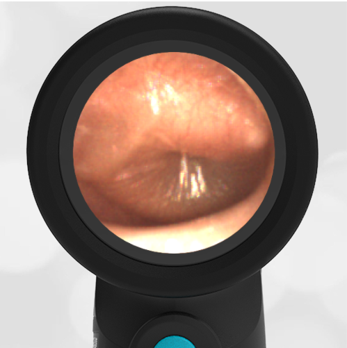
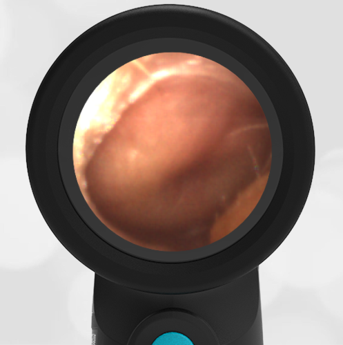
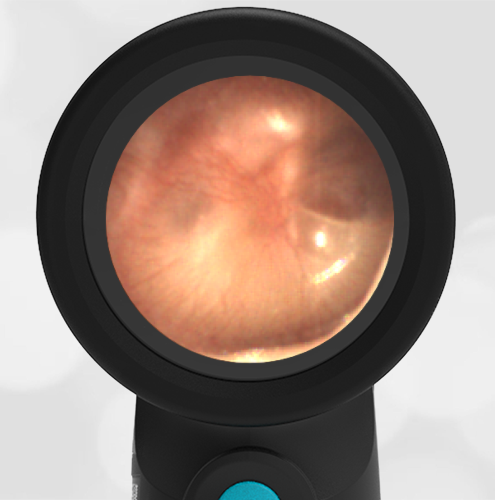
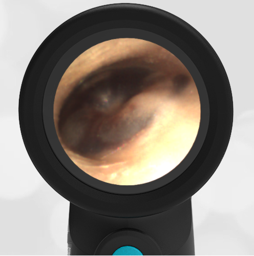
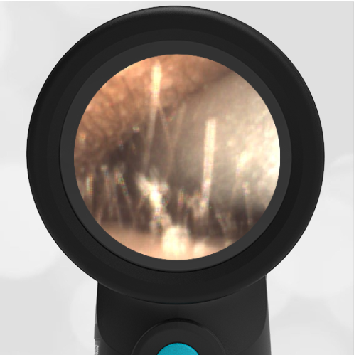
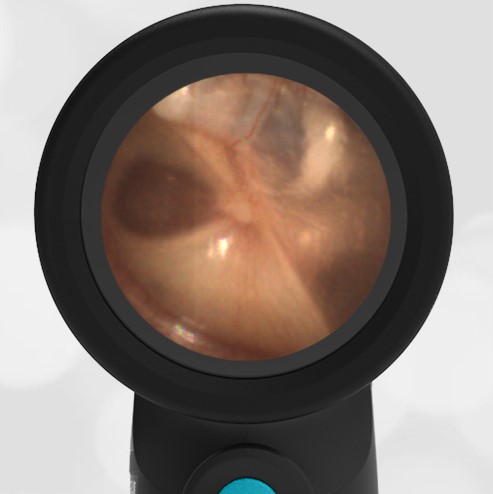
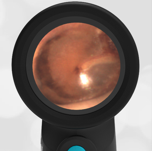
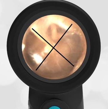
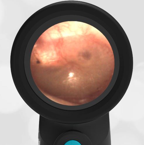
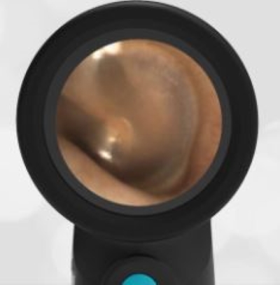
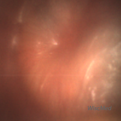
Compare this ear with AOM to a normal ear. It’s easy to tell the difference between the two ears, and also apparent why an ear infection is painful.
-
- Acute Otitis Media
-
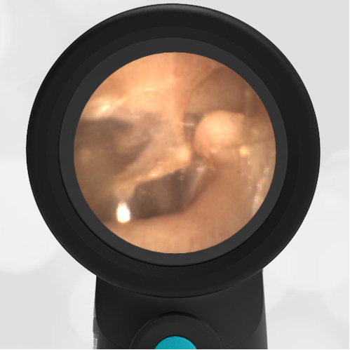
- Normal Ear
Treatment generally involves an oral antibiotic along with topical or systemic pain control. Although the child likely started with a viral infection, when it advances to AOM there is often bacterial co-infection, thus the indication for antibiotics. This Wispr University article tracks the development and resolution of an ear infection through daily images.
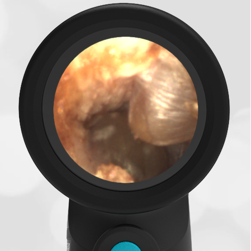
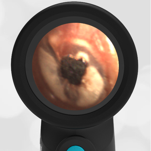
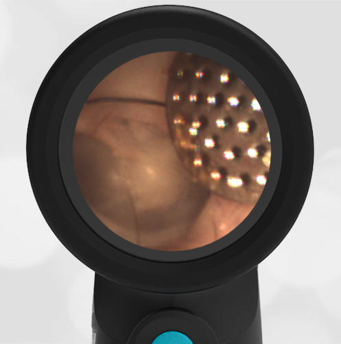
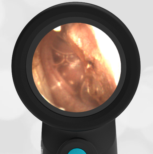
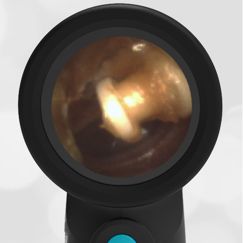
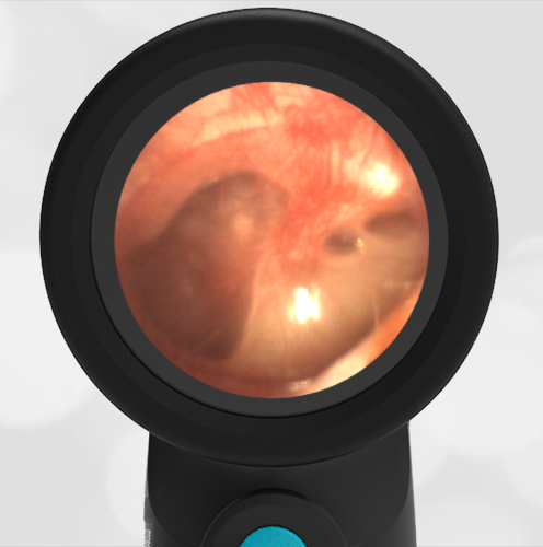
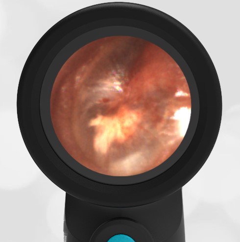
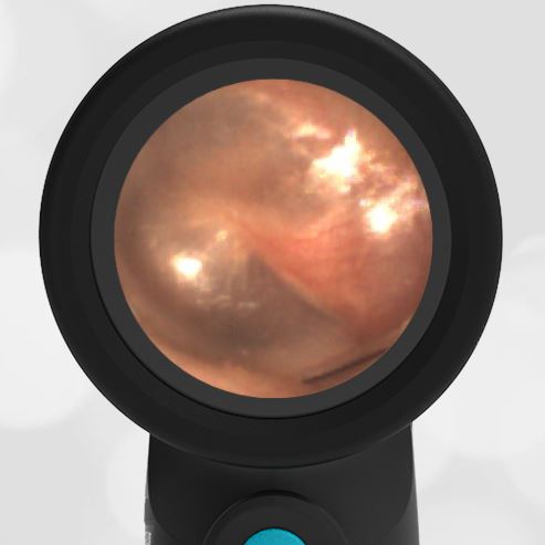
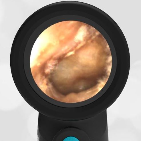
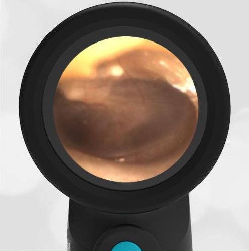
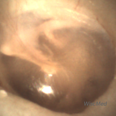
Here is another example of AOM.
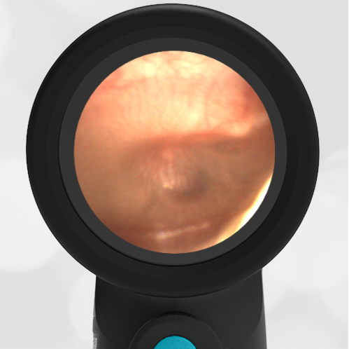
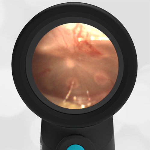
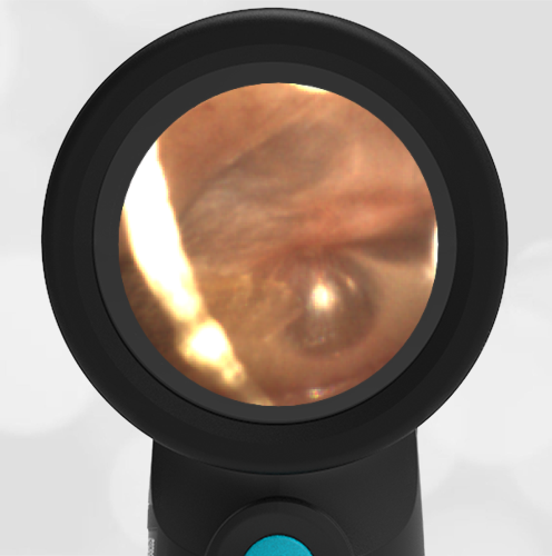
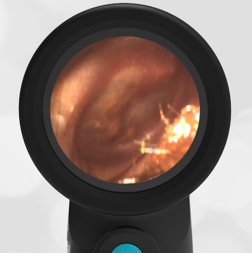
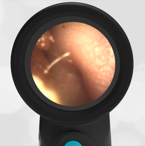
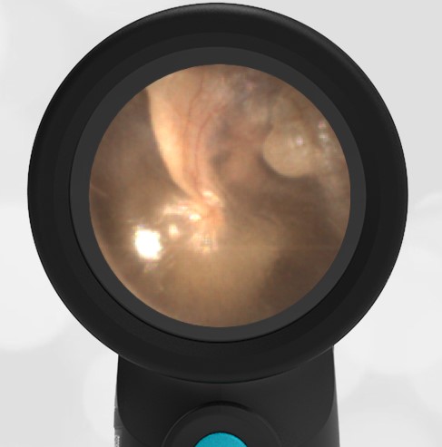
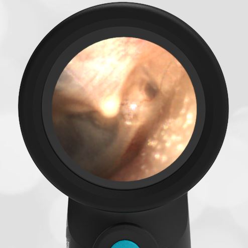
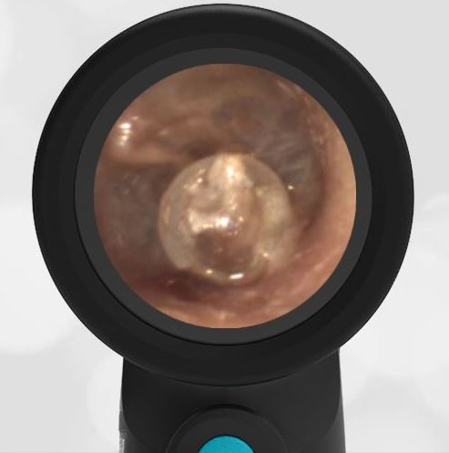
Here is the complete video exam. Note the ability of the Wispr otoscope to navigate the small canal, bypass cerumen (wax) and obtain a diagnostic image.
Complete exam video