CLINICAL CASES
CLINICAL CASES
Using the Wispr Digital Otoscope


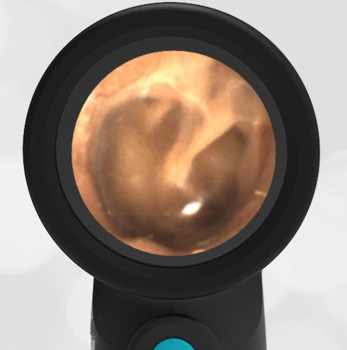
A healthy adult pediatrician visited the Pediatric Academic Societies WiscMed booth for a demonstration of the Wispr digital otoscope. She has no history of ear trauma or infections. She has no ear complaints. The following image of her right ear is obtained with a Wispr digital otoscope.
What unusual feature do you notice about the middle ear bones (ossicles)?

There are three ossicles in the middle ear, the malleus, incus, and stapes. The bones are visible to otoscopy due to the semi-transparent nature of the tympanic membrane (TM). The first bone, the malleus, is easily visible except in cases of anatomic distortion, for example from acute otitis media (AOM). The second bone, the incus, is visible sometimes. The third bone, the stapes, is the deepest of the middle ear bones, and only occasionally can it be seen by otoscopy. The unique observation in this patient is that the inferior portion of the malleus-incus (incudomalleolar) joint is visible. This is unusual as the joint is usually superior to the external ear canal. In this case, the patient has pseudo retraction of the pars flaccida portion of the TM. Because of this, there is an unusual opportunity to appreciate the joint. Here is the video of the complete ear exam:
Complete exam video

