Ear Ventilation Tube
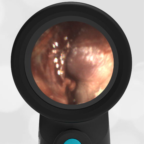
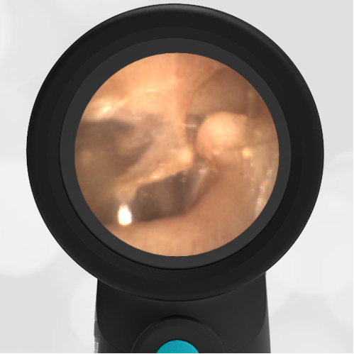
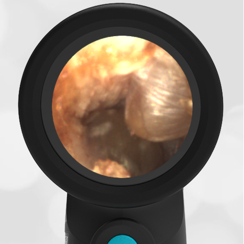
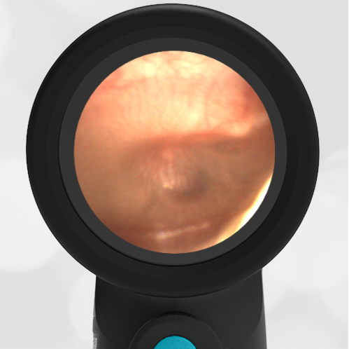
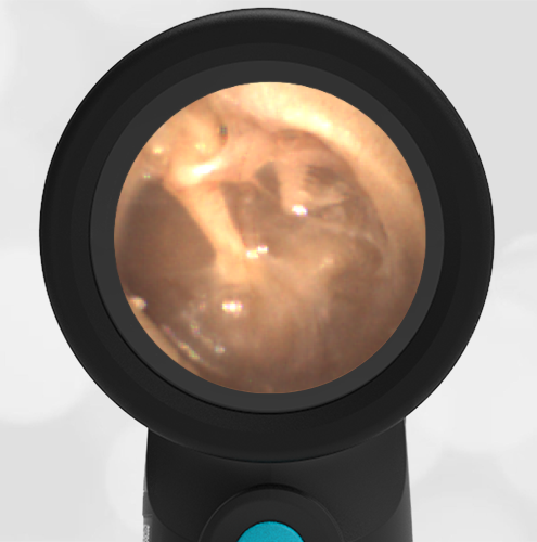
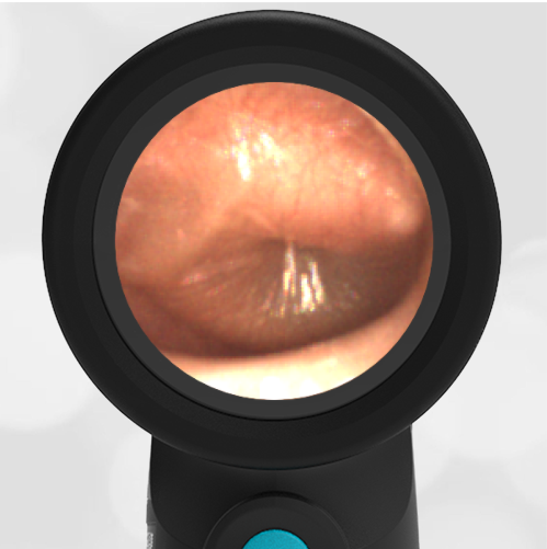
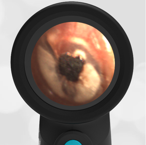
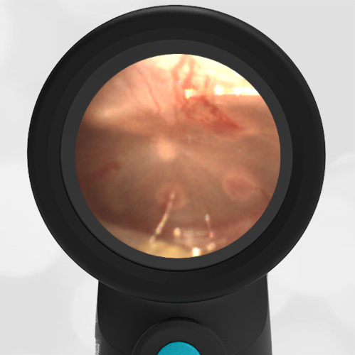
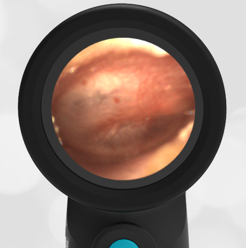
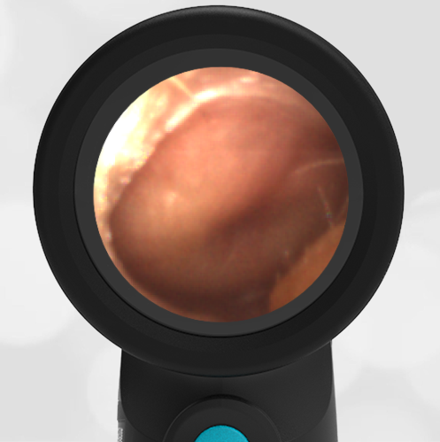
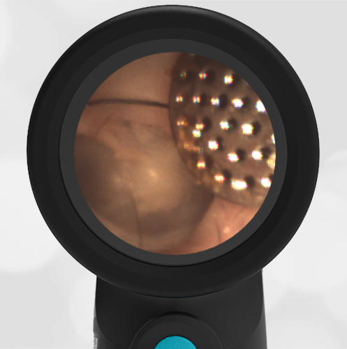
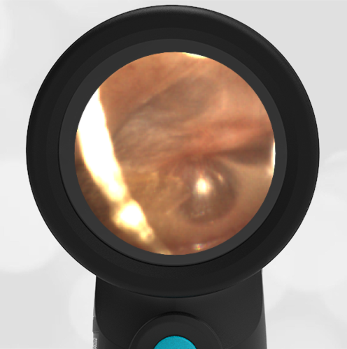
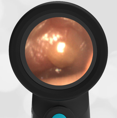
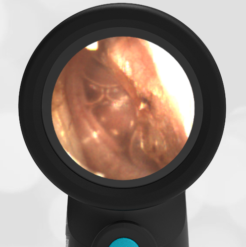
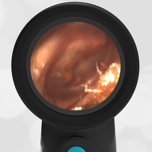
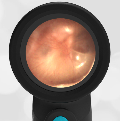
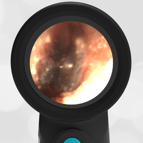
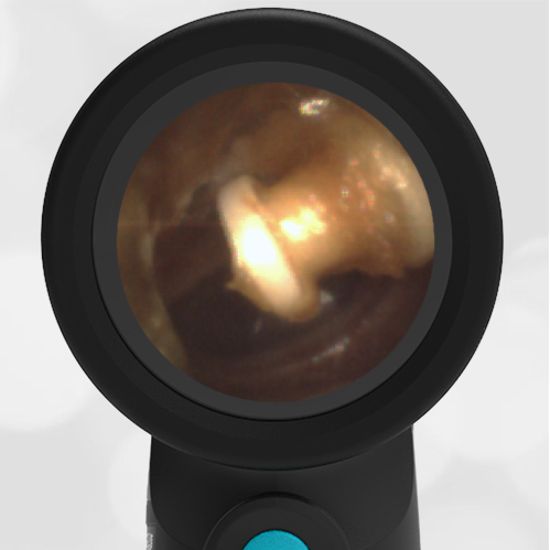
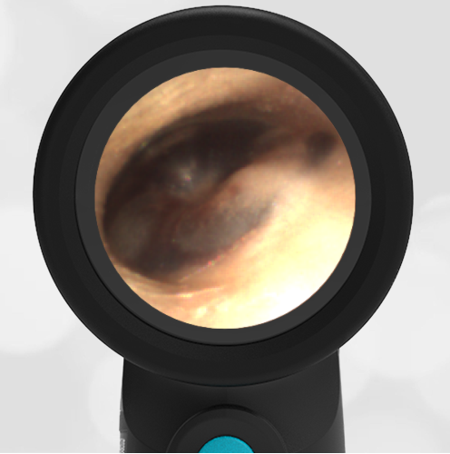
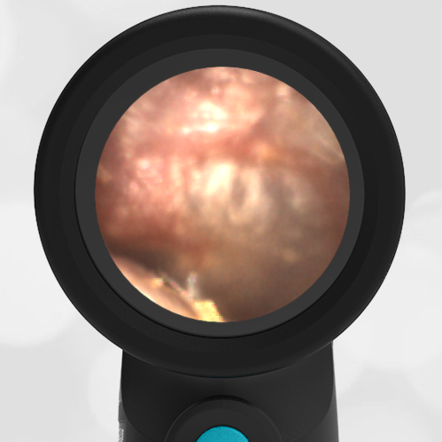
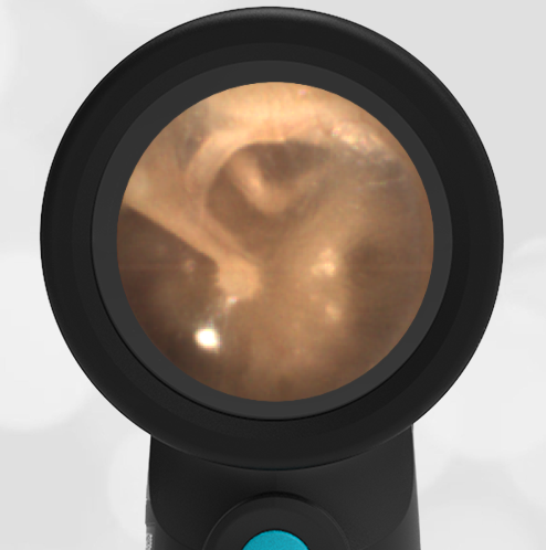
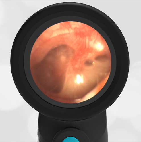
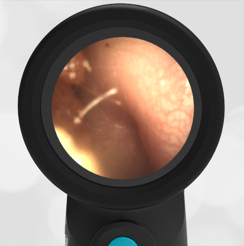
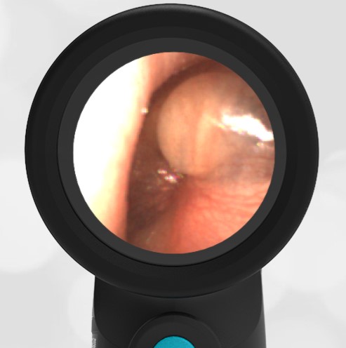
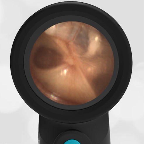
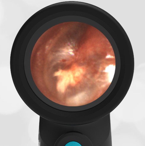
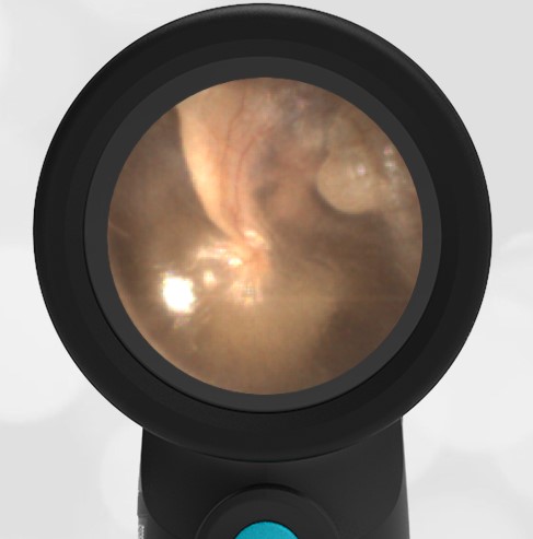
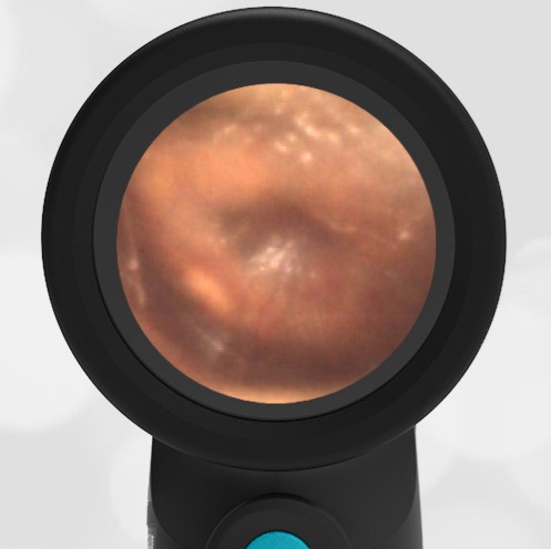
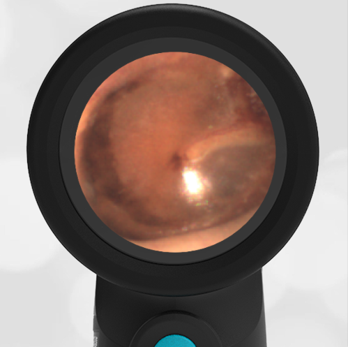
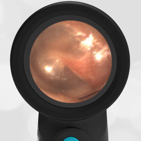
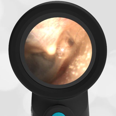
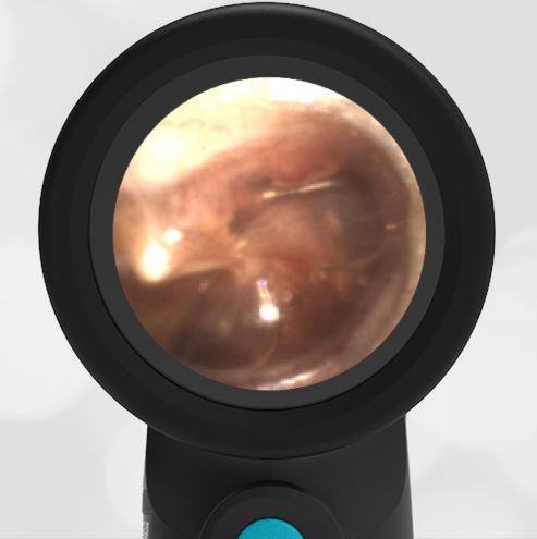
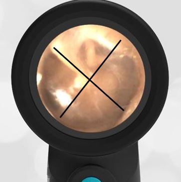
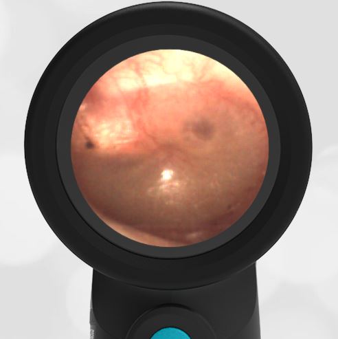
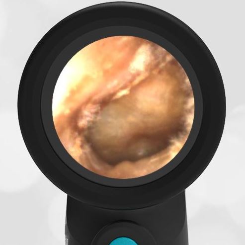
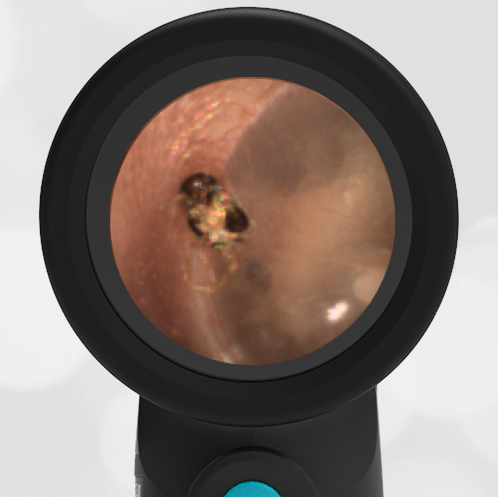
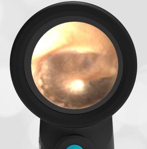
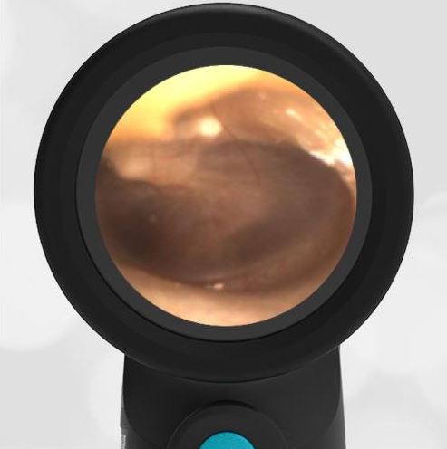
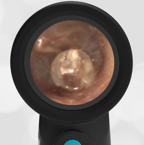
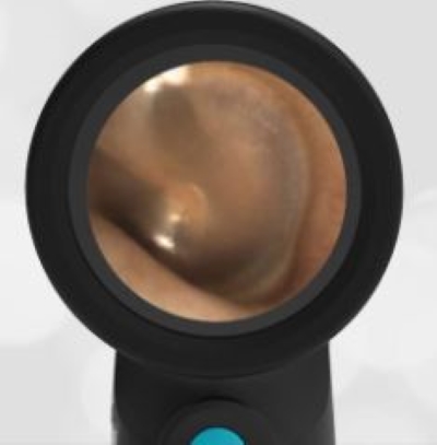
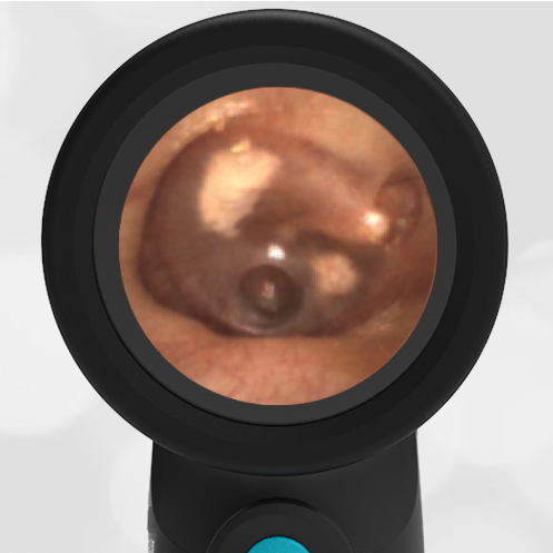
A 6-year-old male presents to the pediatric clinic for a well-child check. His mother does not have any concerns today. The child has a history of acute otitis media and had ventilation (ear) tubes placed about 3 months ago. The following image of the right ear was obtained.
What action needs to be taken today?
The patient has a patent, well-placed ventilation tube without evidence of infection. No action is necessary.
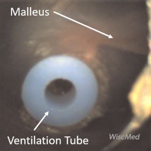
Ventilation tubes, also known as “ear tubes” and tympanostomy tubes, are commonly placed for cases of recurrent acute otitis media (AOM) and less commonly for chronic middle ear effusions (MEE). The tubes are placed in the anterior-inferior quadrant of the TM. This location is directly opposite the location of the Eustachian tube orifice in the middle ear. Tympanostomy tubes provide equalization of pressure across the TM. They essentially temporarily replace the job of a poorly functioning Eustachian tube. Small children are susceptible to Eustachian tube dysfunction because their anatomy causes the tube to be relatively horizontal, causing drainage challenges. As children grow, the Eustachian tube becomes more vertical, allowing proper drainage to the posterior nasopharynx. This partially explains why ear infections are so much more common in children than adults. In this case, the TM appears normal as does the malleus bone. The ventilation tube is well-placed and its annulus appears patent.
The ventilation tubes eventually fall out on their own, typically in 6 to 12 months. The tympanic membrane generally heals with no long-term hearing consequences. Occasionally, there is evidence of prior ear tub placement. Often, there is myringosclerosis of the TM which has no clinical significance.


