Tympanic Perforation
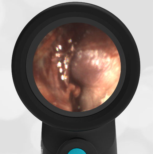
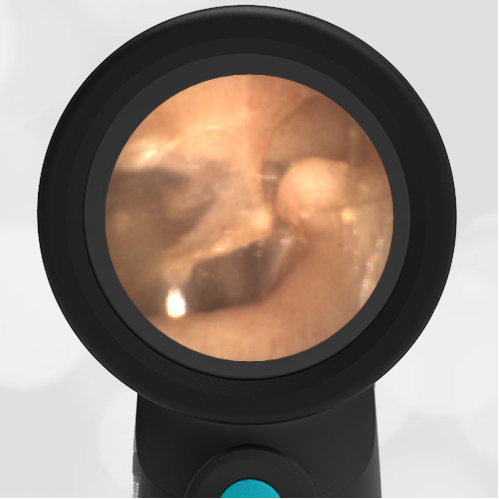
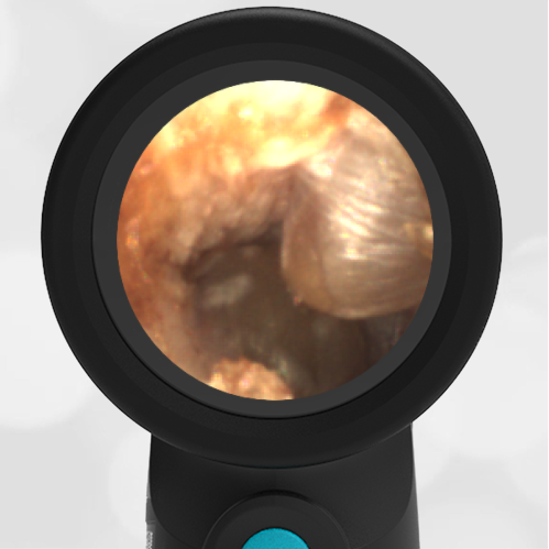
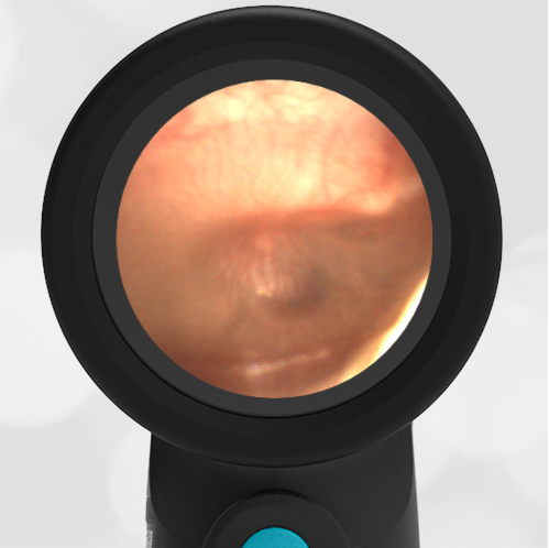
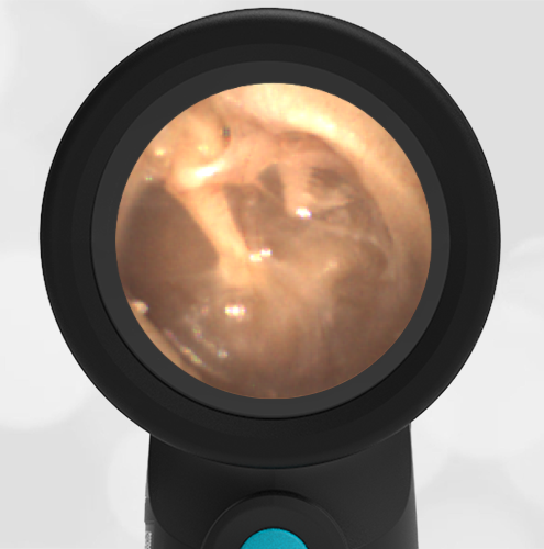
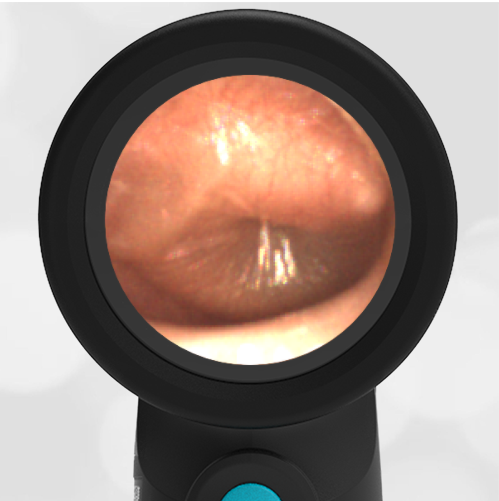
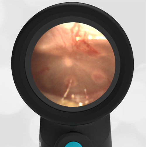
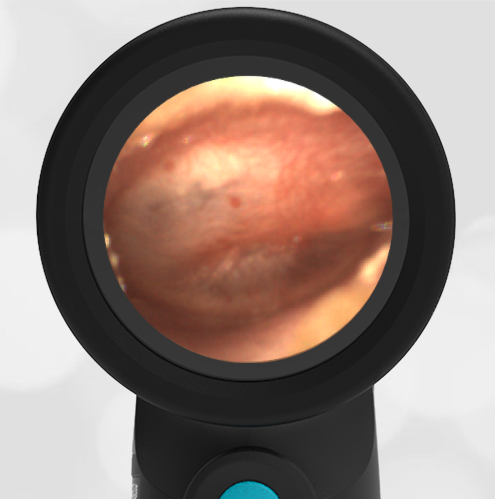
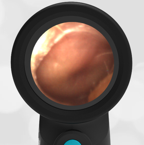
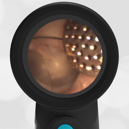
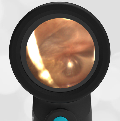
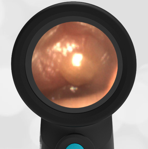
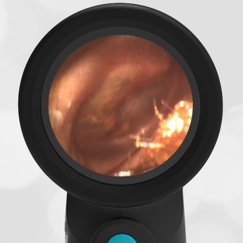
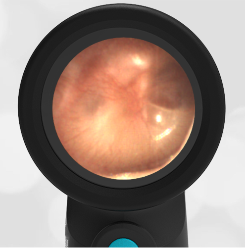
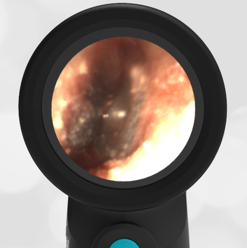
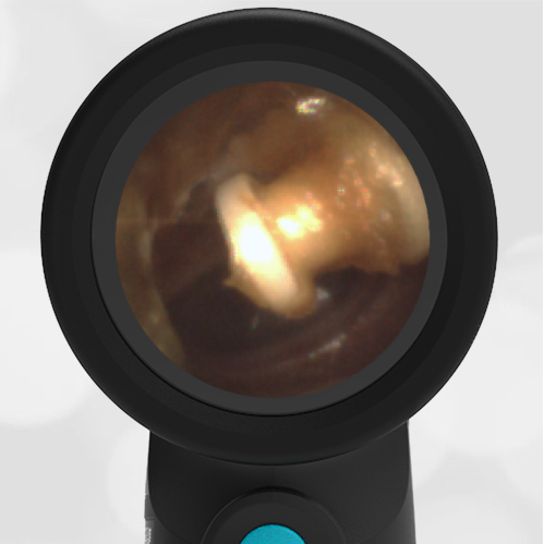
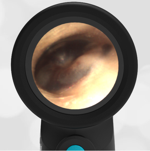
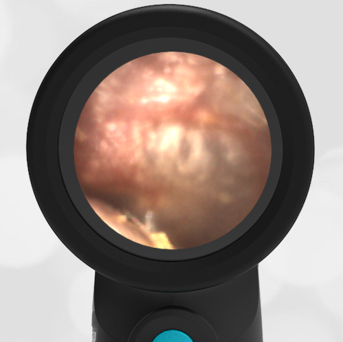
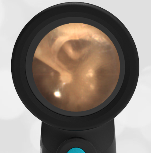
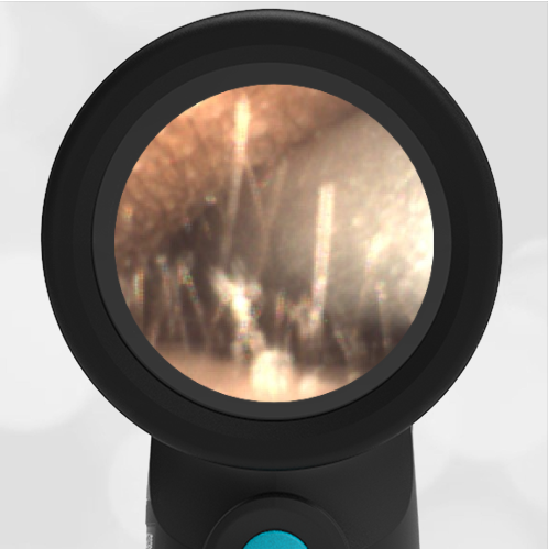
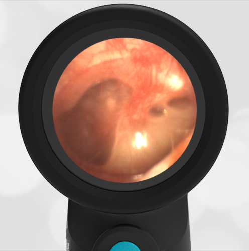
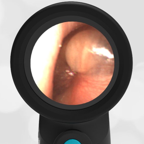
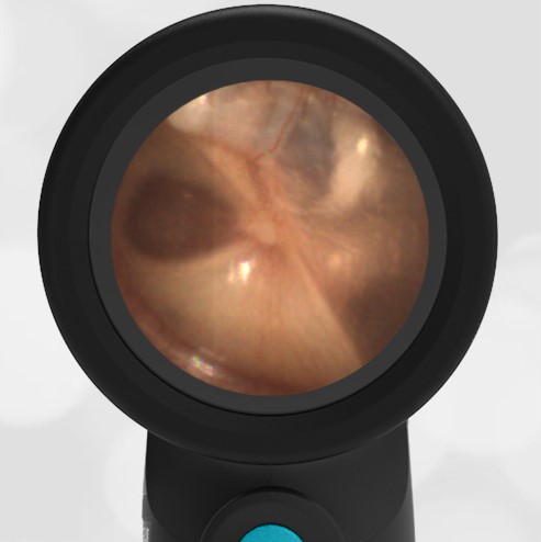
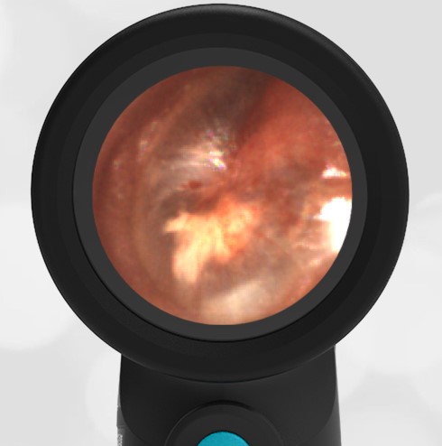
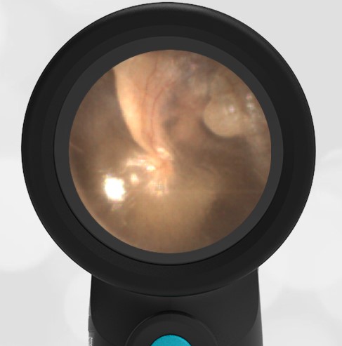
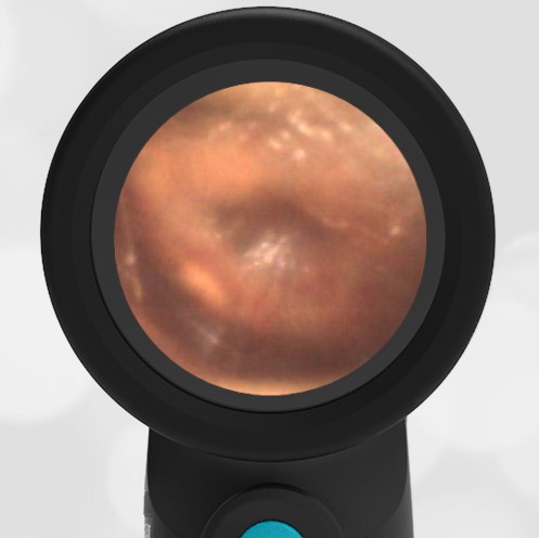
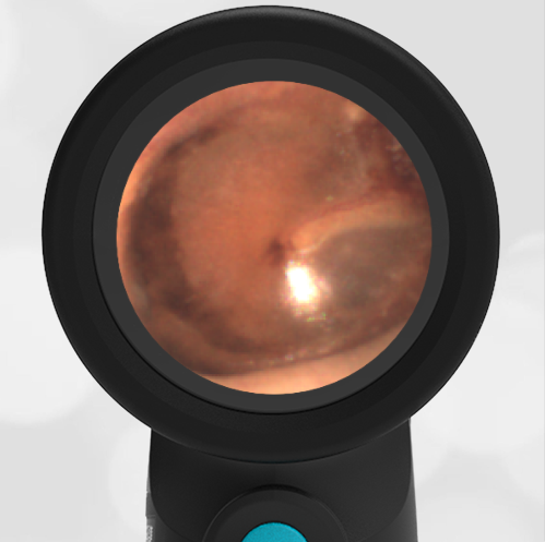
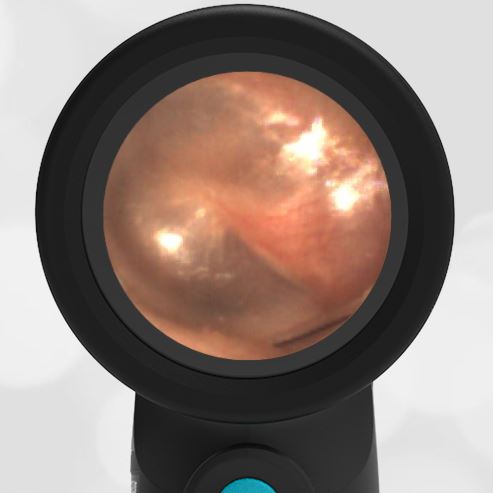
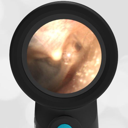
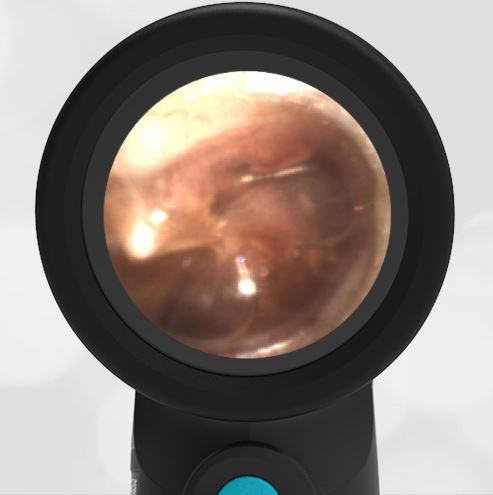
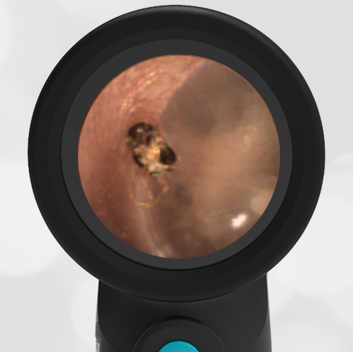
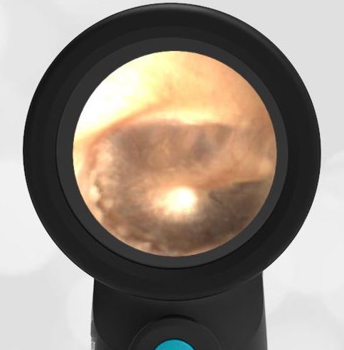
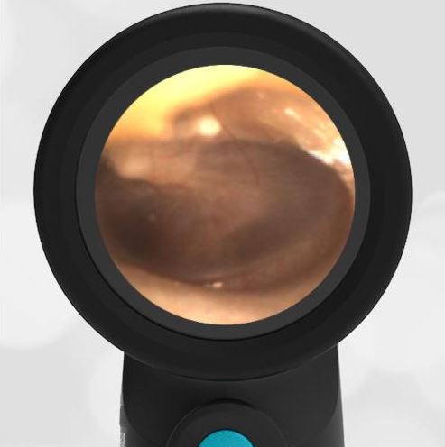
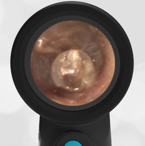
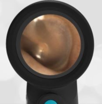
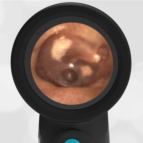
A 6-year-old boy with a history of recurrent acute otitis media (AOM) presents with drainage from his left ear for one day. His father reports the child used to have frequent ear infections and had tubes placed a couple of years ago. Since that time, he has not had any ear infections although his hearing is somewhat diminished in the left ear compared to the right ear. He is supposed to be following up with his ENT for “some procedure”. His Wispr exam reveals this image and the likely indication for this procedure.
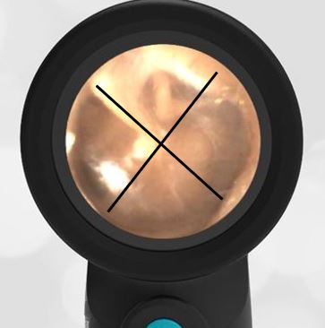
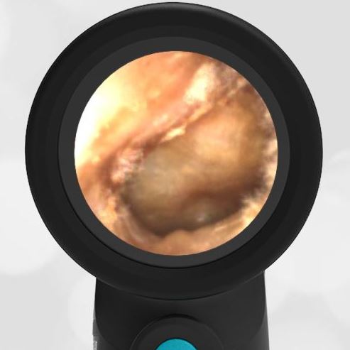
This child has a persistent tympanic perforation following the removal of his tympanostomy tubes (TM).
Additionally, there is mild otorrhea consistent with AOM. Upon further questioning, the child’s dad recalls a minor procedure last year when the TM tubes were indeed removed from both ears.
Persistent perforation is a known complication following TM tube placement, although the reported rates vary widely from 2% (Grommet type) to 24% (T-tubes). If the tubes are not expelled naturally, removal may be indicated after 18 to 24 months. Since most perforations close spontaneously, they may be observed up to a year before intervention with tympanoplasty or a variety of patching techniques. However, if left untreated perforations may cause conductive hearing loss and place the child at risk for recurrent infection. At his most recent ENT appointment, it was felt that he may require a myringoplasty due to the persistent perforation and diminished hearing.
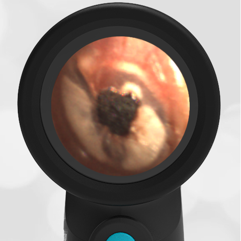
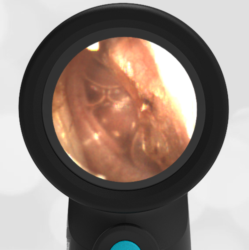
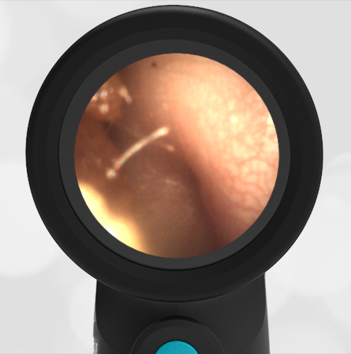
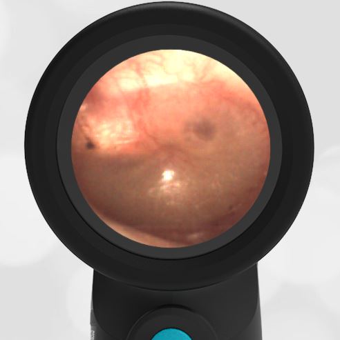
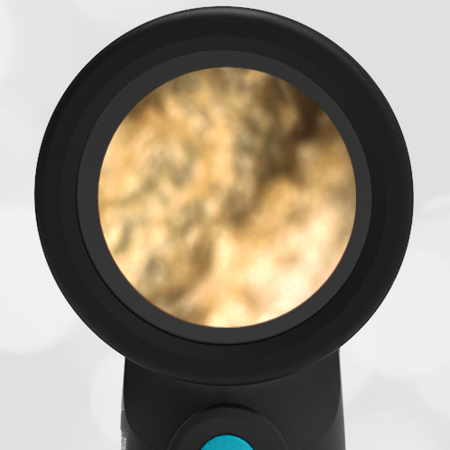
Incidentally, the astute clinician will note the presence of the “Black Lab Sign” on the child’s Wispr exam. Father notes the family did in fact have a very loving Labrador retriever that frequently slept with the patient.
Here is the video of the exam:
Complete exam video


