Recent Acute Otitis Media – March 28, 2024
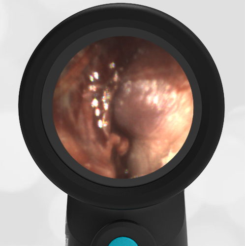
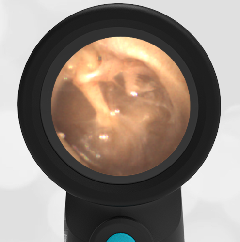
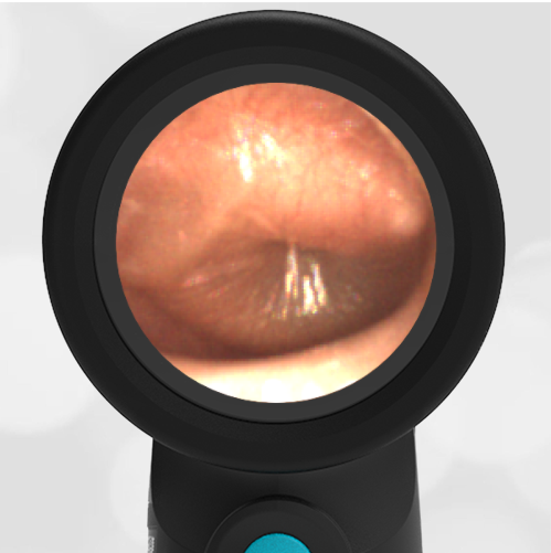
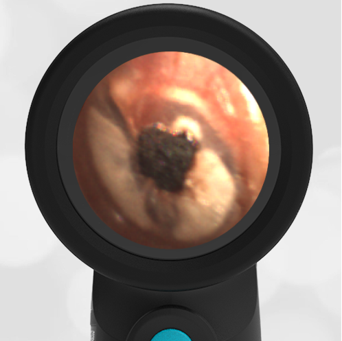
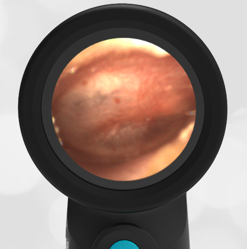
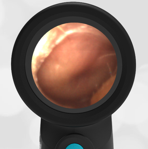
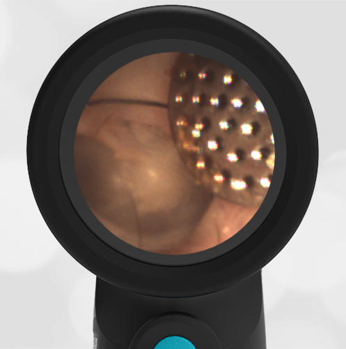
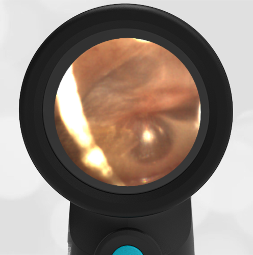
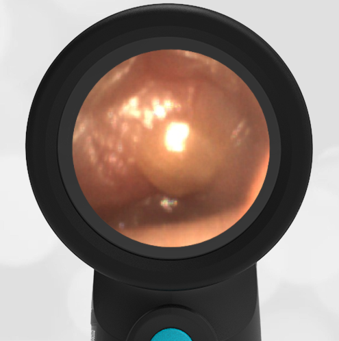
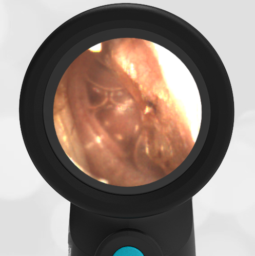
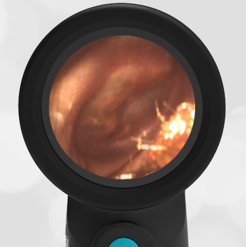
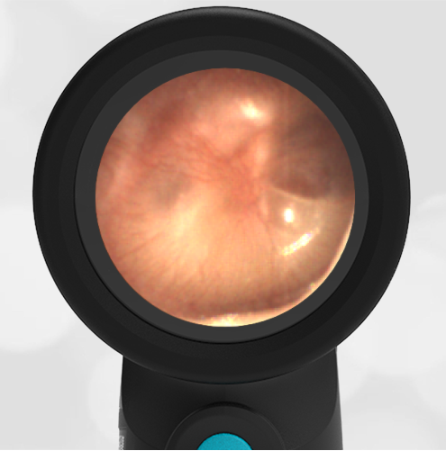
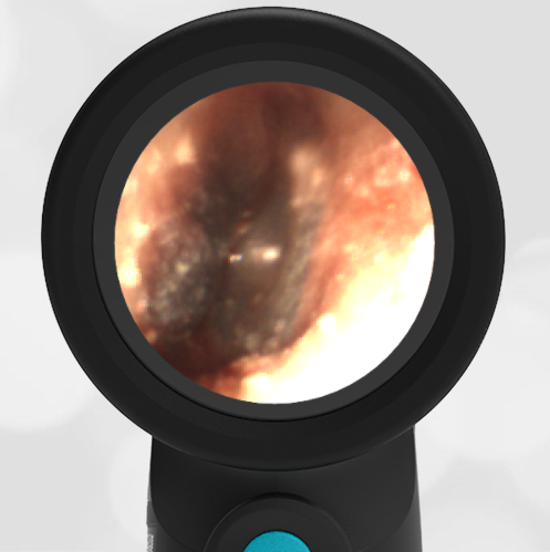
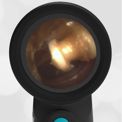
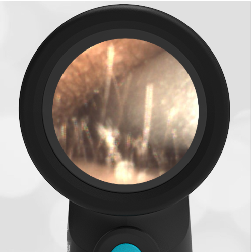
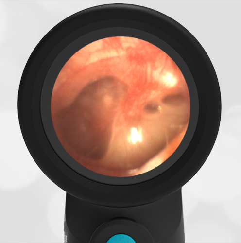
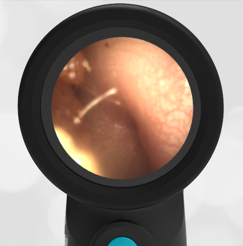
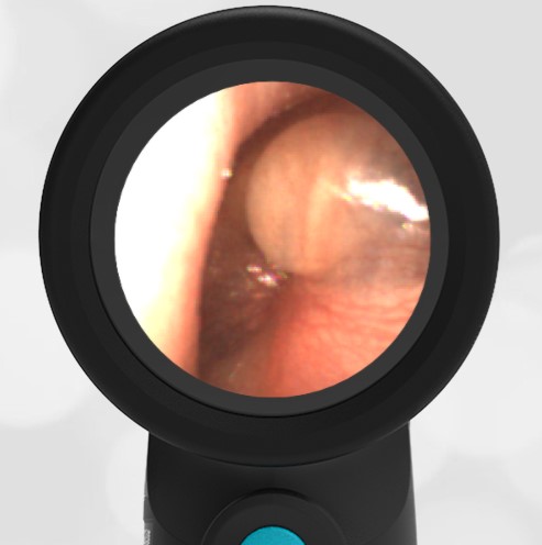
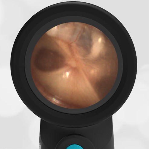
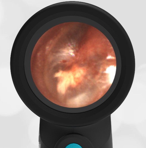
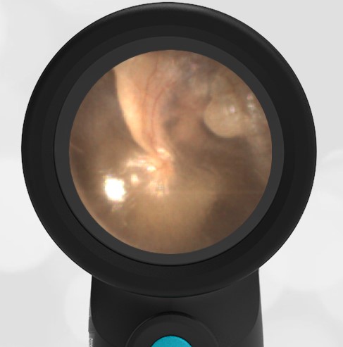
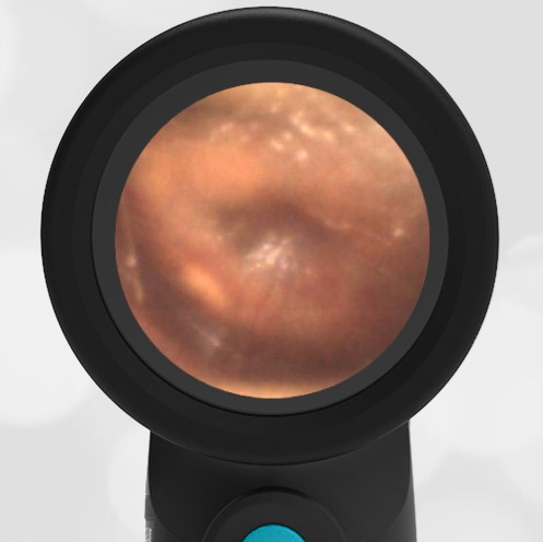
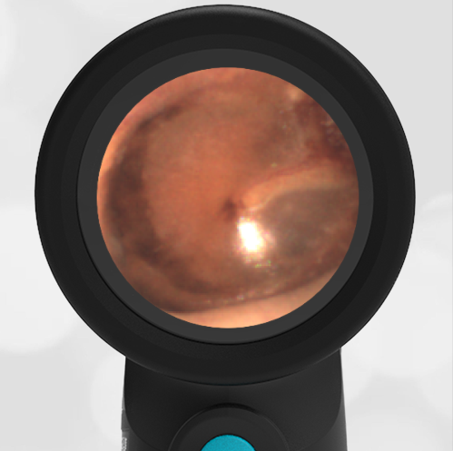
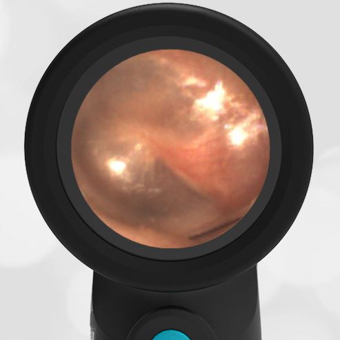
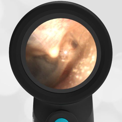
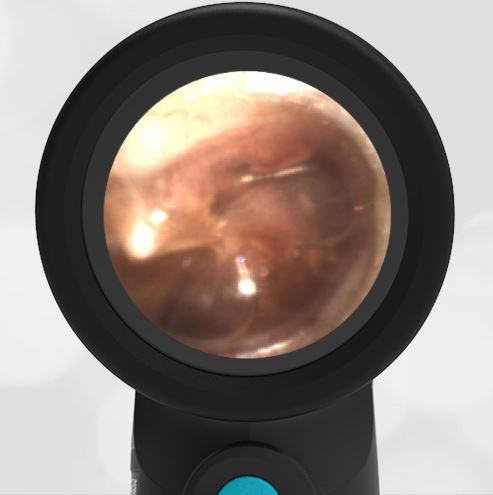
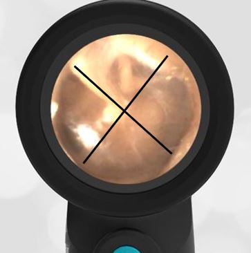
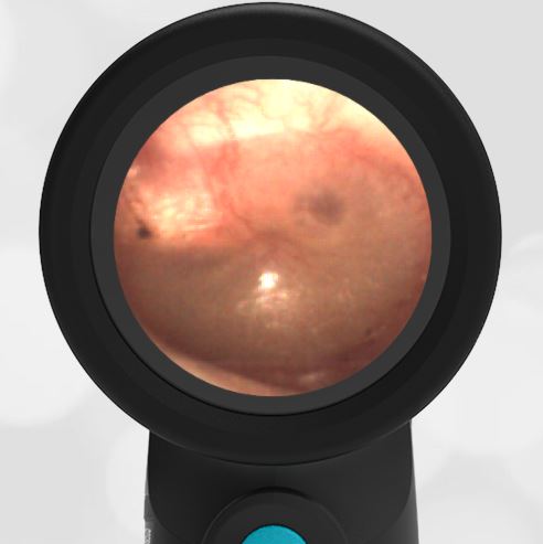
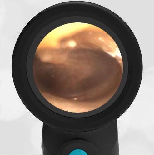
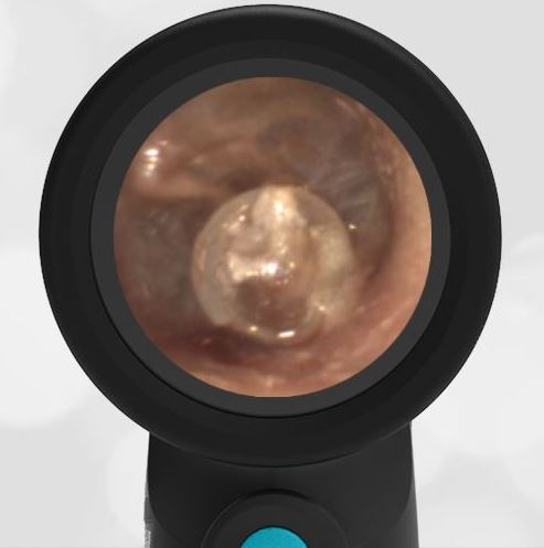
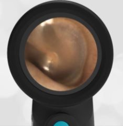
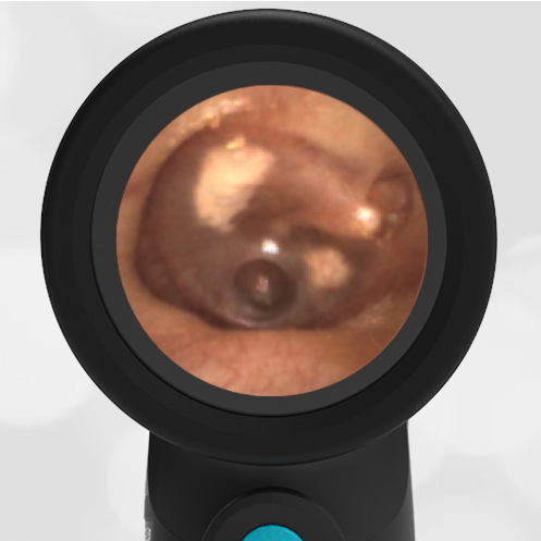
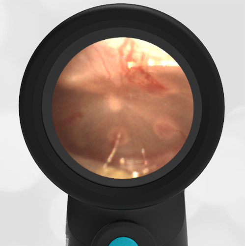
A 3-year-old presents to the emergency department (ED) for congestion, cough, and shortness of breath. Her parents report this is her second or third “cold” since starting daycare two months ago. The child is afebrile and generally well-appearing. She has mild wheezing on auscultation that resolves with albuterol. Her Wispr digital otoscope exam is shown.
Which of the following can be inferred from the tympanic membrane (TM) findings?
- She recently had acute otitis media (AOM).
- She has had multiple ear infections and tympanostomy tubes (TT).
- She previously had a ruptured tympanic membrane (TM, “ear drum”).
- Given her history, an urgent, but not emergent consult to otolaryngology (ENT) is indicated.
Answer: A. She recently had acute otitis media (AOM).
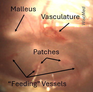
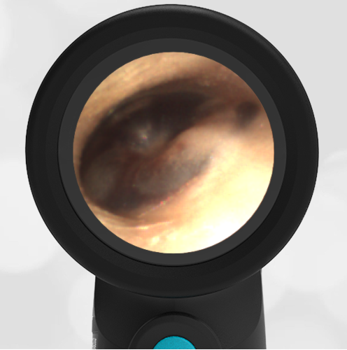
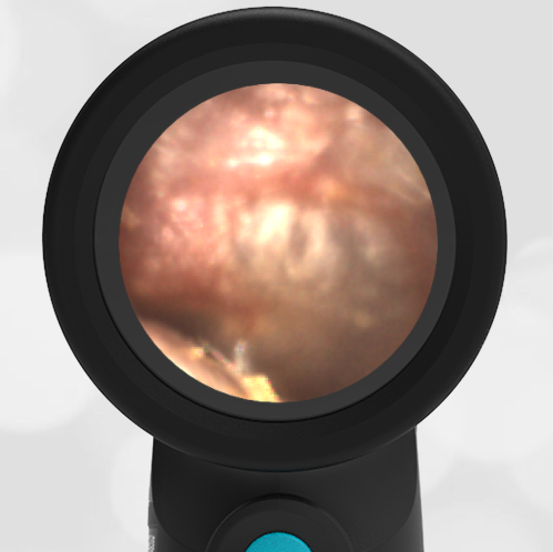
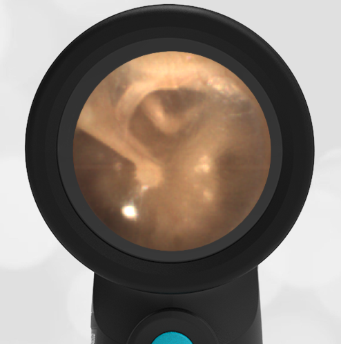
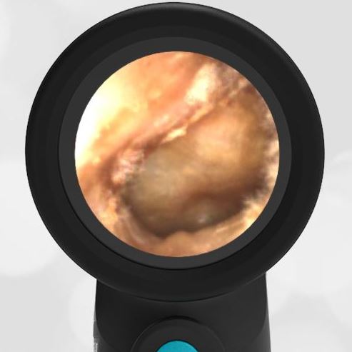
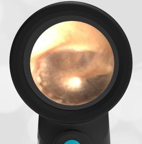
The child’s Wispr exam demonstrates several small circular patches along the periphery of the tympanic membrane (TM, “ear drum”). These patches of mucopurulent material are on the “inside” of the TM and may appear during the recovery phase of acute otitis media. In the image, small blood vessels involved in healing are also easily appreciated. Dr. Michael Poole in his book, “Otitis, the Expert’s Diagnostic Guide” refers to these small blood vessels as sentinel vessels or feeding vessels (chapter 9.2). Tympanosclerosis (also termed myringosclerosis) are areas of thickened TM resulting from recurrent AOM, injury, or tympanostomy tubes. Tympanosclerosis is not present here. AOM recovery patches are transitory, while tympanosclerosis is permanent and appears as asymmetric white plaques that lack vasculature. There is no indication of a past perforation. There is no indication for a referral to ENT.
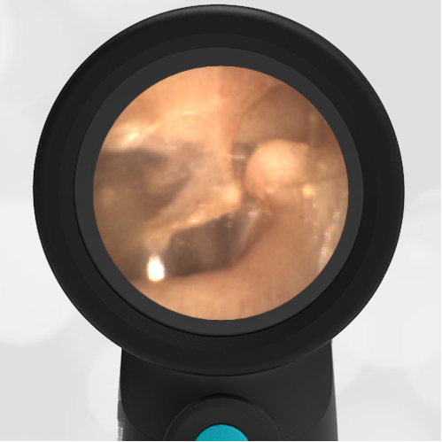
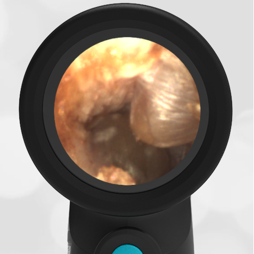
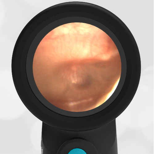
Here is the complete video exam