Retraction Pocket
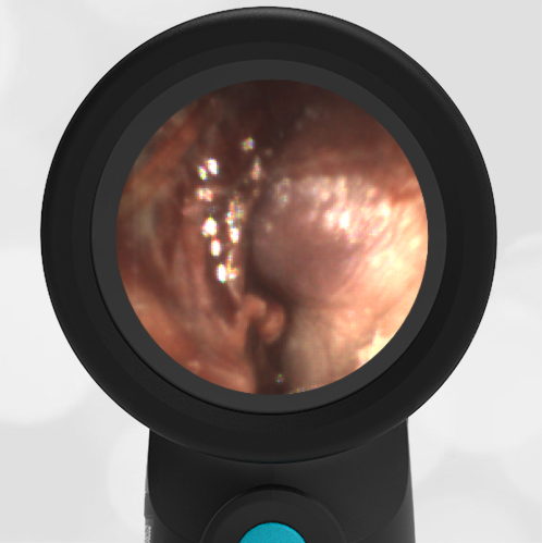
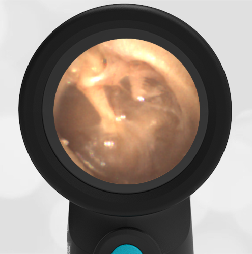
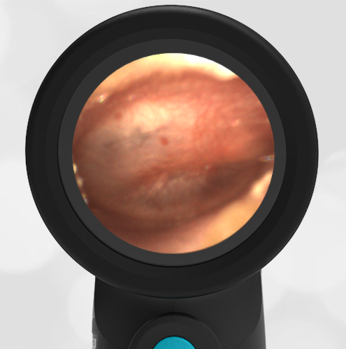
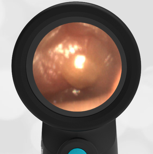
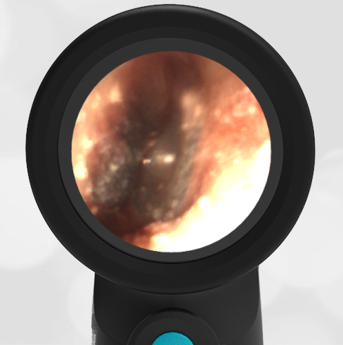
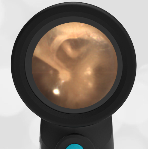
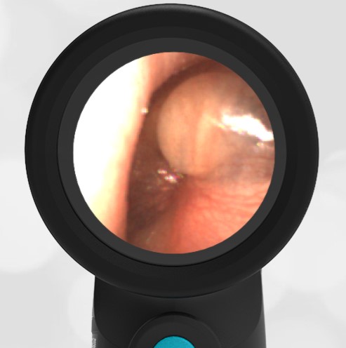
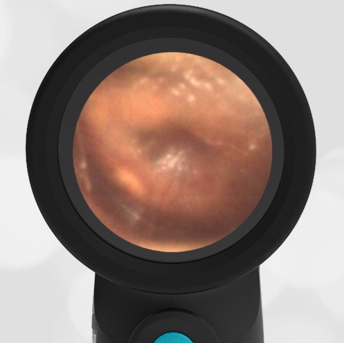
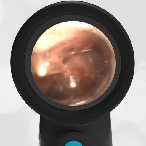
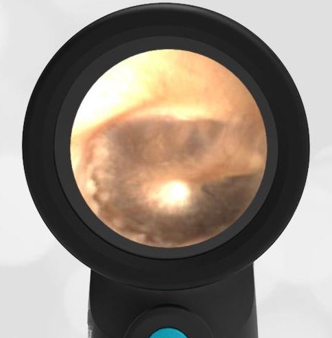
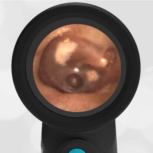
A 5-year-old female presents with complaints of congestion and cough. Her mother reports “at least a month” of symptoms. The child has a history of frequent ear infections and completed a course of amoxicillin three weeks earlier. In the ED she is afebrile and well-appearing. She has no ear complaints. Her Wispr exam of the left ear is shown. Upon review of the images with mother, she asks “How did that hole get there?”
The best answer to provide the child’s mother is:
- The child likely perforated her tympanic membrane (TM)
- The “hole” is a result of TM tube placement
- The finding is associated with negative pressure in the middle ear space
- “I don’t know”
The correct answer is “3.” The “hole” is actually retraction pocket.
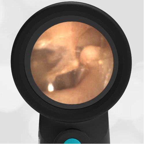
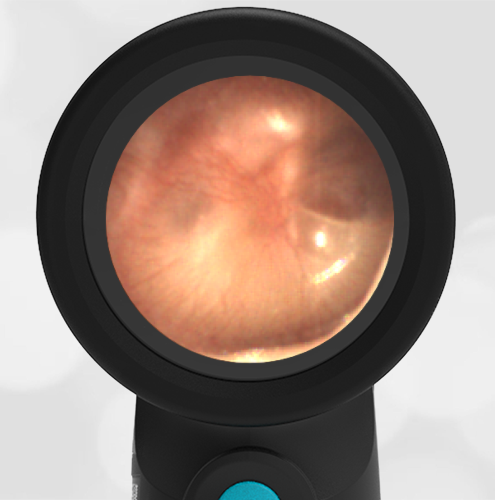
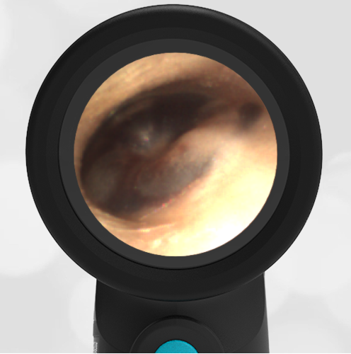
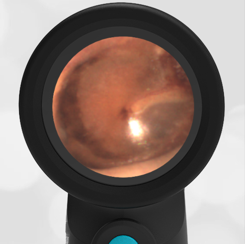
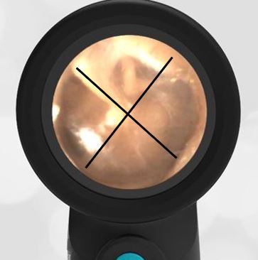
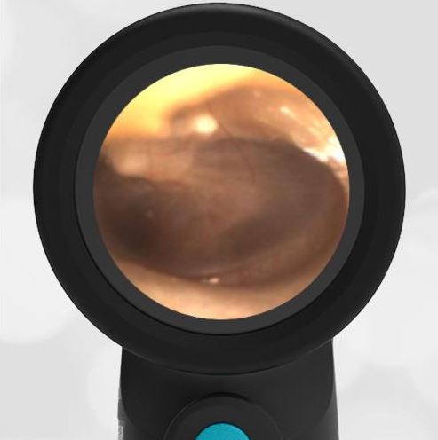
While it is easy to see how the mother (and clinician) could mistake the dark oval area in the posterior-superior quadrant of the TM for a hole (rupture), this finding is an example of a mild retraction pocket (RP).

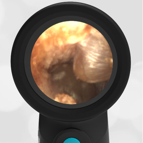
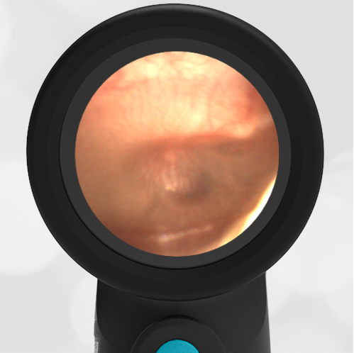
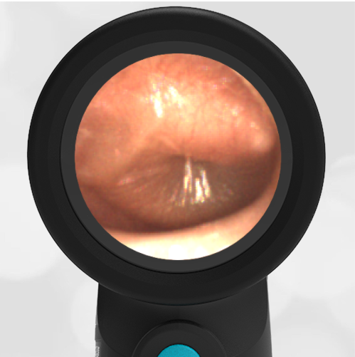
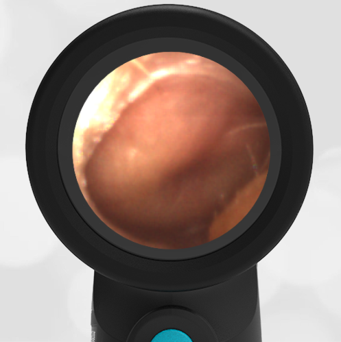
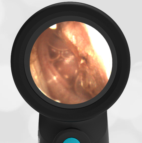
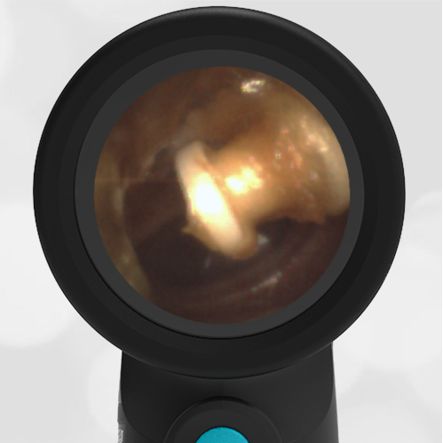
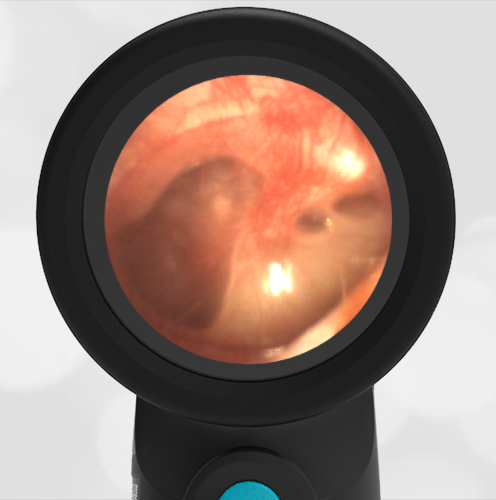
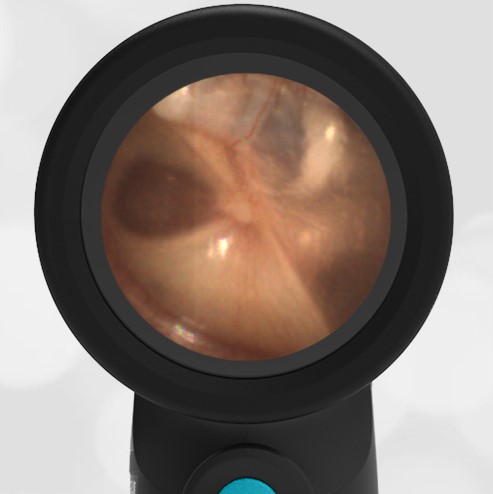
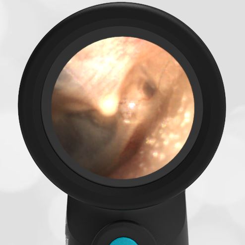
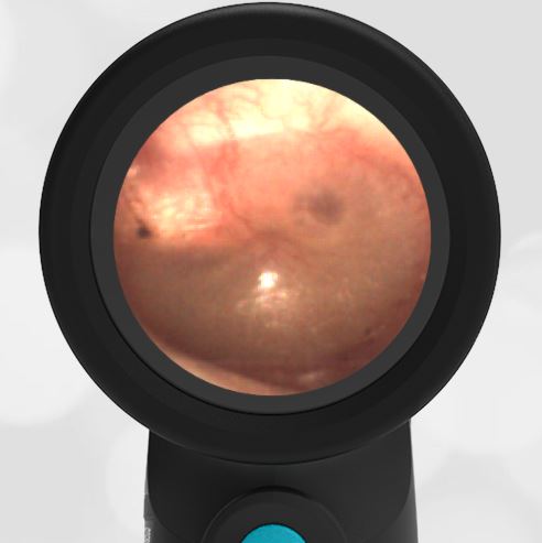
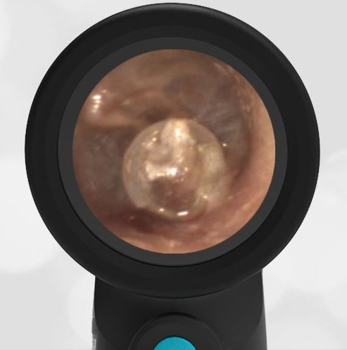
A retraction pocket is a localized invagination of the TM most often forming in the pars flaccida region (but not in this case). While the exact etiology is under debate, it is likely brought on by a combination of factors including chronic negative pressure and inflammation within the middle ear space. Thus, Eustacian tube dysfunction and recurrent or chronic otitis media are felt to play a role in its formation which may explain why they are much more common in children than adults. Here is an example of an eardrum retraction in an adult.
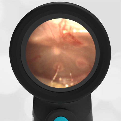
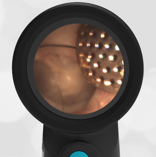
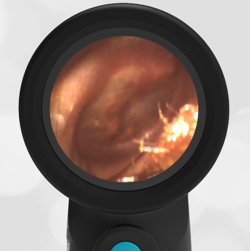
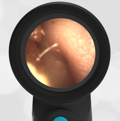
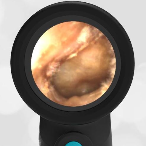
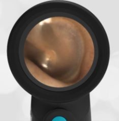
The malleus ossicle (bone) is a prominent feature seen on otoscopy. It is the first bone in the chain-of-three that translates sound waves to the middle ear.
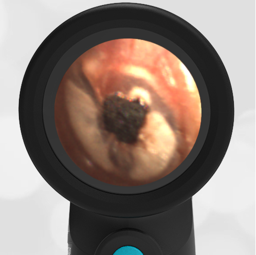
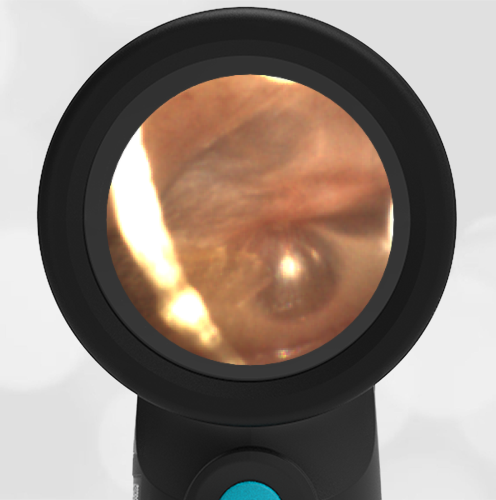
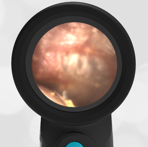
While small RPs often resolve spontaneously and remain asymptomatic, there is the possibility of progression. In these instances, otorrhea, otalgia, erosion of ossicles, cholesteatoma formation, and hearing loss may occur. Since optimal management (also under debate) may involve surgical excision and tympanoplasty, follow-up with ENT is warranted to ensure the resolution of deeper or symptomatic RPs.
Here is the complete video exam:
Complete exam video
Urik M.; tedla M.; Hurnik P. Pathogenesis of Retraction Pocket of the Tympanic Membrane—A Narrative Review Medicina 2021, 57(5), 425; https://doi.org/10.3390/medicina57050425
Bunne, M.; Falk, B.; Magnuson, B.; Hellstrom, M. Variability of Eustachian tube function: Comparison of ears with retraction dis-ease and normal ears. Laryngoscope 2000, 110, 1389–1395.