CLINICAL CASES
CLINICAL CASES
Using the Wispr Digital Otoscope


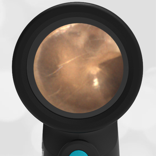
A 15-year-old female presents to the pediatric clinic for a routine sports exam. She is well and has no complaints. This image of her right ear is obtained. She is a curious student and asks you to identify and explain the structures seen in the image.
What structures would you choose to identify and explain?
One of the benefits of youth is that anatomy is often pristine, as in the case of this beautiful right ear image.

The ear drum (tympanic membrane, TM), separates the outer ear from the middle ear space. It is the only structure that is “outside.” The other features are “behind” the translucent eardrum in the middle ear space. Think of the eardrum as being a cloudy layer of taught saran wrap. The surface responds to sound waves by vibrating. This vibration must be turned into a signal that our brain can process. The job of translating the vibration to the inner ear is accomplished by a chain of three tiny bones; the malleus, incus, and stapes. These are often referred to as the hammer, anvil, and stirrup. The malleus is directly attached to the inner surface of the eardrum. As the eardrum vibrates, the malleus moves with the vibration. The malleus is connected to the incus and the incus to the stapes. Through these connections, the tiny movement of the eardrum is mechanically amplified and delivered to the inner ear. In the inner ear (not visible), this movement is converted to a fluid action that moves tiny hairs which create the nerve signal to the brain. It’s a beautifully engineered system. Also visible in this image is the chorda tympani nerve. It runs behind the malleus and in front of the incus. This nerve has nothing to do with the ear, it simply borrows the middle ear space to get “where it needs to go.” It provides taste sensation from part of the tongue along with secretory signals. It’s unusual to be able to see this nerve so clearly.

