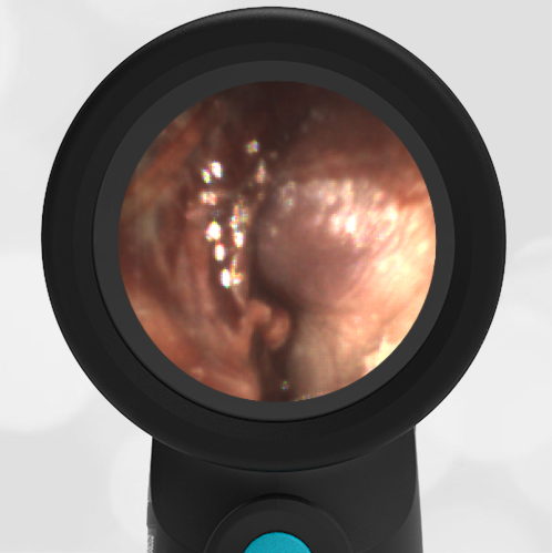
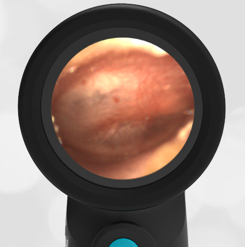
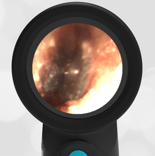
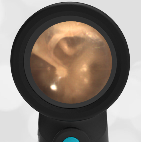
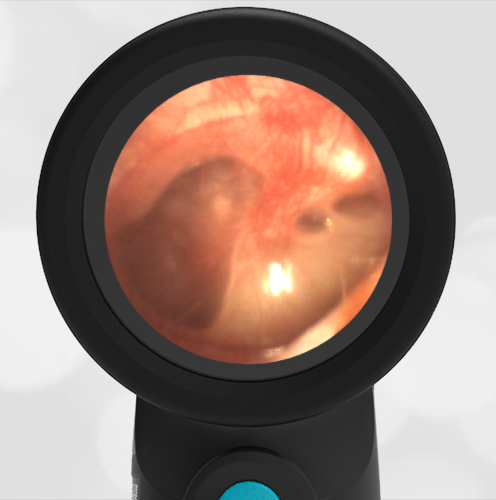
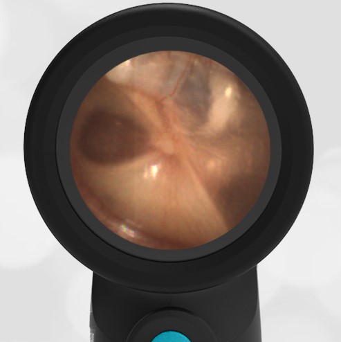
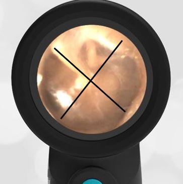
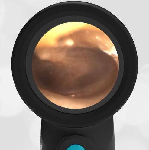
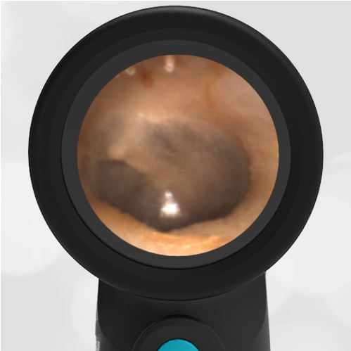
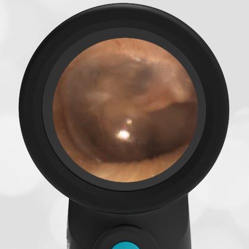
Which Ear?
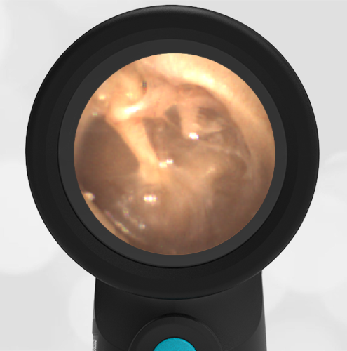
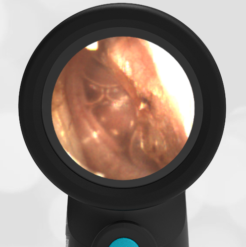
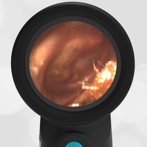
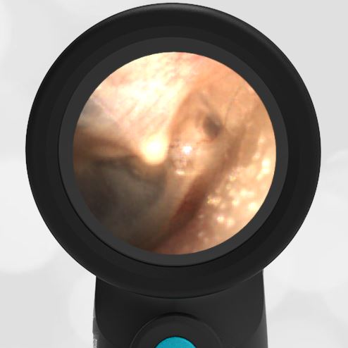
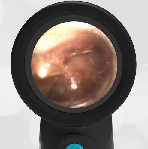
A healthy 23-year-old female presents for a routine sports physical exam. This image is obtained Wispr Digital Otoscope. Unfortunately, the examiner forgot to make note of which ear the image is from for the medical record. How can you tell which ear the image is from? This is one of our favorite questions to ask learners because the answer is simple and useful.
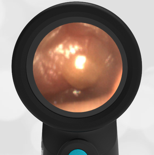
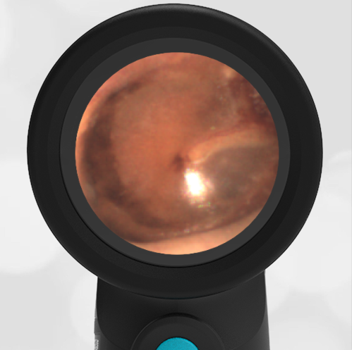
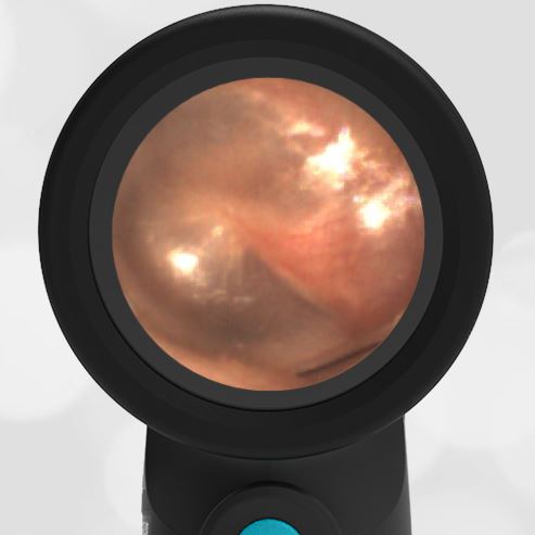
This image is from the right ear.
It’s easy to tell because of the orientation of the malleus bone. The malleus points like an arrow towards the face of the patient.

A prominent feature of the ear exam is the presence of the malleus bone. This is the first of three bones in the middle ear that communicates the movements of the eardrum, from sound waves to the inner ear. The formal names of the three bones are malleus, incus, and stapes. These are commonly referred to as “hammer, anvil, and stirrup.” Conveniently, the malleus is like an orientation compass for the examiner in an ear with normal anatomy.
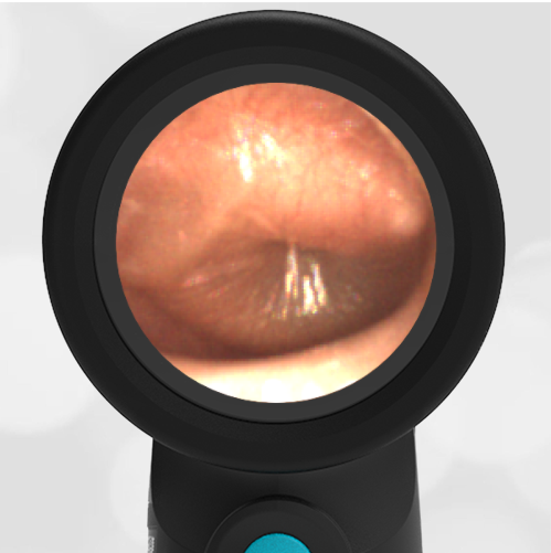
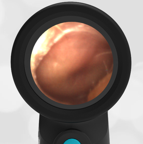
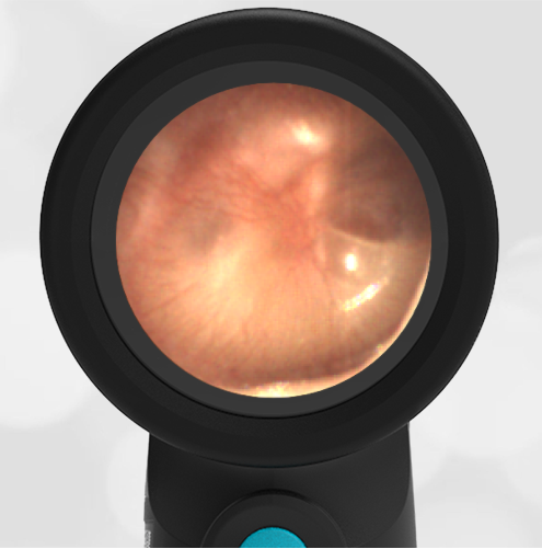
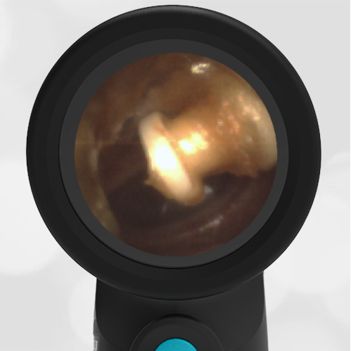
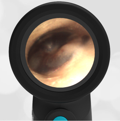
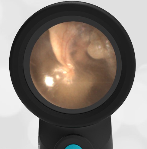
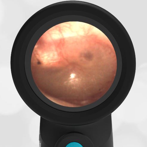
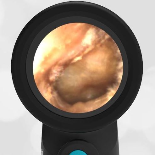
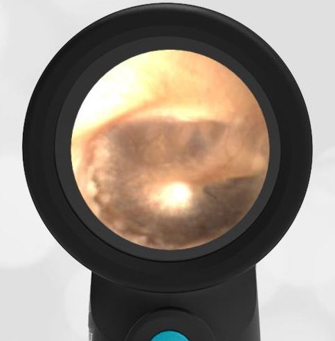
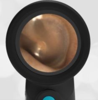
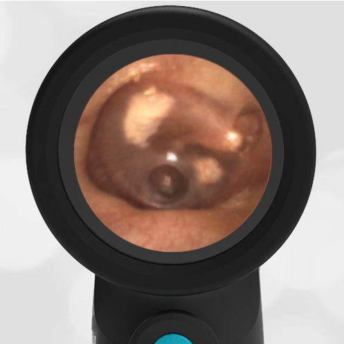
Compare the right ear and the left ear taken from the same patient.
-
- Left Ear
-
- Right Ear