CLINICAL CASES
CLINICAL CASES
Using the Wispr Digital Otoscope


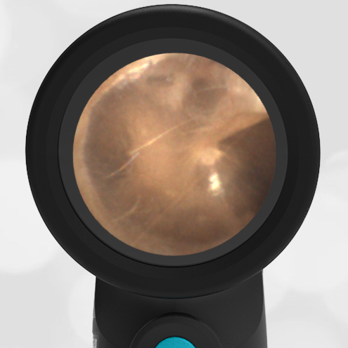
A healthy 25-year-old male with no complaints sees his primary care physician for a routine checkup. This image of the patient’s ear is obtained. He asks you to explain what he is seeing in the image.
What structures can you outline for this curious patient?

This is a nice example of a healthy and normal tympanic membrane (TM). The most obvious structures are the three bones of the middle ear, the malleus, incus and stapes. These three bones are actually “behind” the tympanic membrane. They are visible because the TM is a thin and partially translucent membrane. Their purpose is to translate and amplify the vibration of the eardrum to the inner ear. The malleus is connected to the incus superior to what is visible through the TM. It is unusual to be able to see the most distant bone, the stapes, as it tends to be further away from the TM.

In addition to the bones of the middle ear, the Chorda Tympani nerve is also visible. This nerve runs behind the malleus and in front of the incus. It has nothing to do with sensation or motor to the ear. It borrows this convenient middle ear space as it makes its way to the tongue to provide taste sensation and innervation to the sublingual and submandibular glands in the mouth. The Eustachian tube shadow is the opening of the Eustachian tube in the middle ear. This tube connects the middle ear to the posterior nasopharynx. Its job is to equalize pressure on both sides of the TM. Its function is familiar to us as it allows our ears to “pop” during the descent of an airplane.

