Acute Otitis Media (AOM)
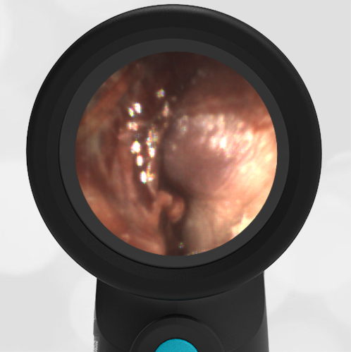
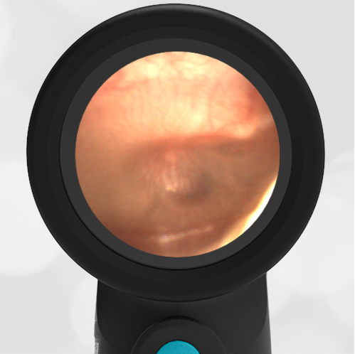
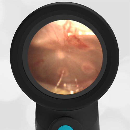
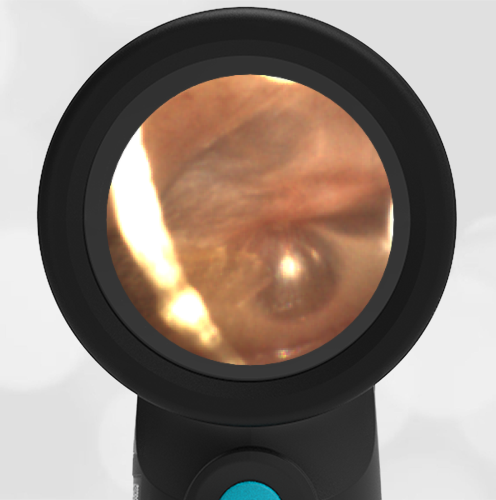
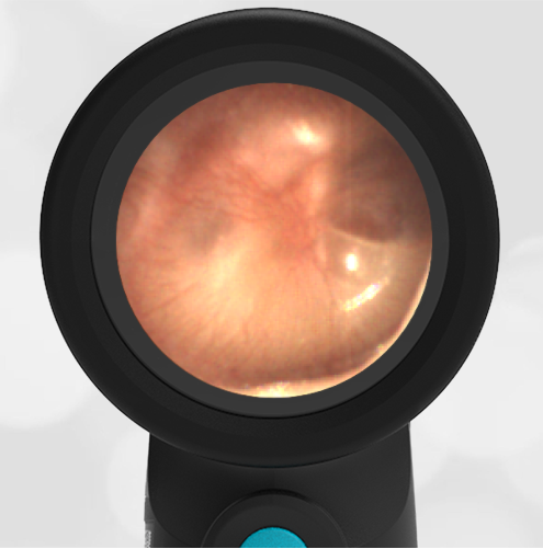
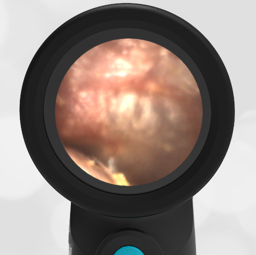
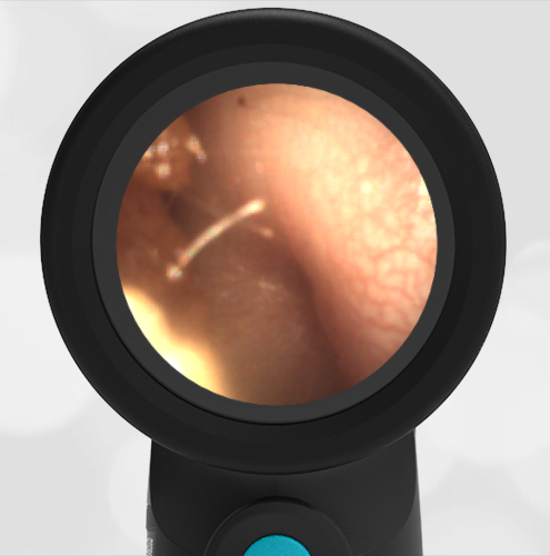
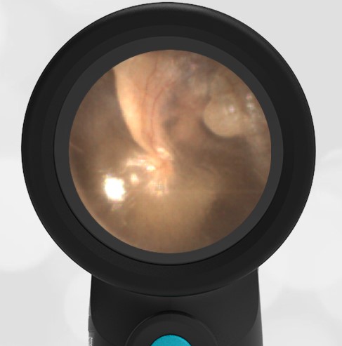
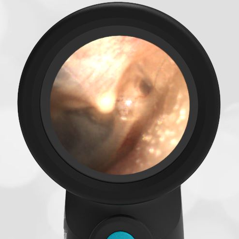
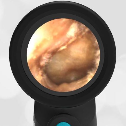
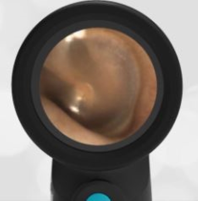
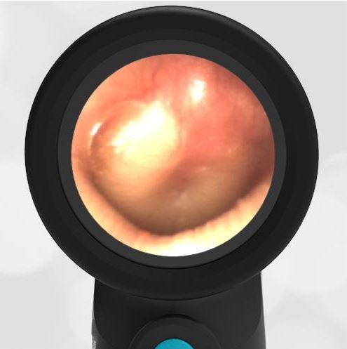
A 10-month-old previously healthy male is brought in by his mother with concern for fever and fussiness. The child continues to take nourishment and has wet diapers. On exam, he is alert, bright-eyed, drooling with a brisk capillary refill. Rhinorrhea is noted, and breath sounds are normal. Ear exam reveals this image.
This child has acute otitis media (AOM).
The tympanic membrane is bulging (donut-shaped) with erythema and loss of bony landmarks. An exam of the other ear reveals the same condition. The child was started on antibiotics for bilateral acute otitis media and did well.
Acute Otitis Media (AOM)
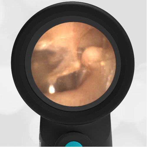
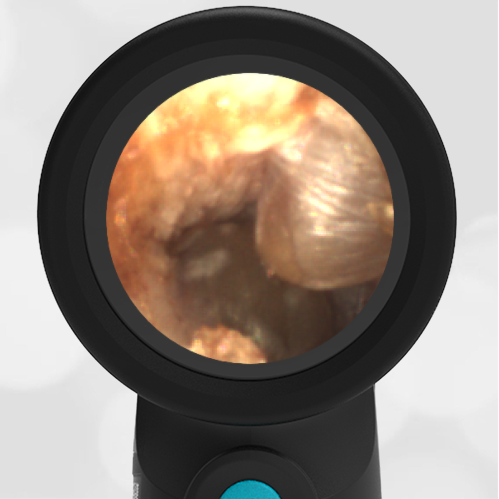
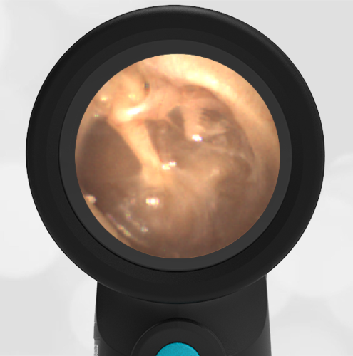
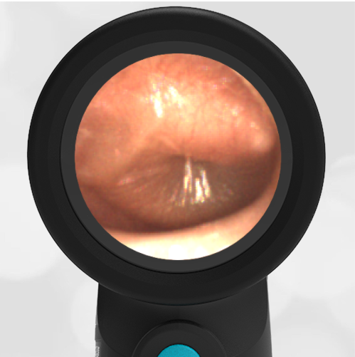
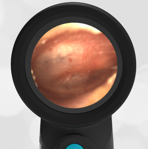
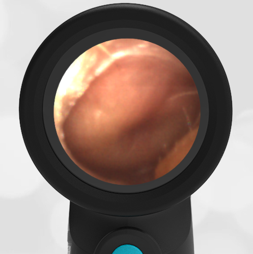
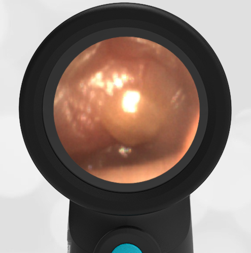
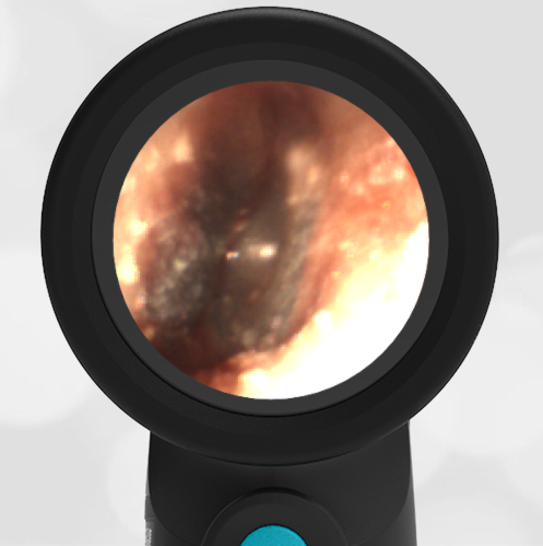
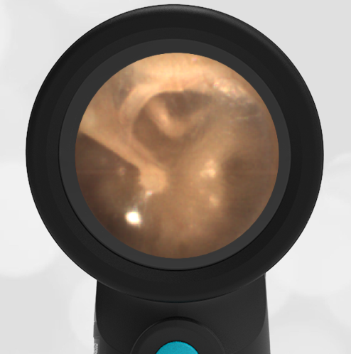
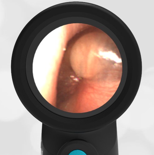
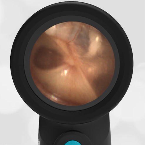
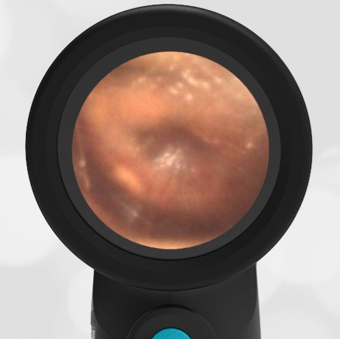
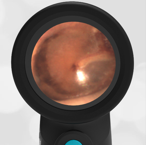
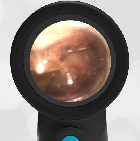
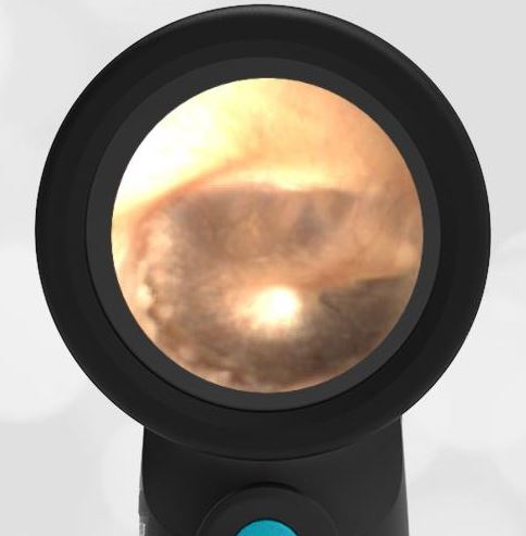
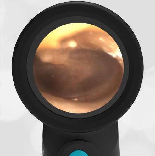
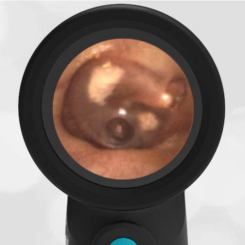
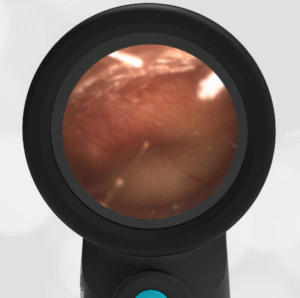
Compare the difference between middle ear effusion (MEE) and AOM.
-
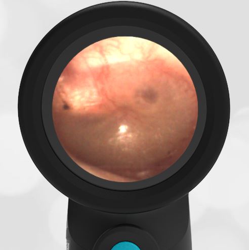
- Middle Ear Effusion (MEE)
-
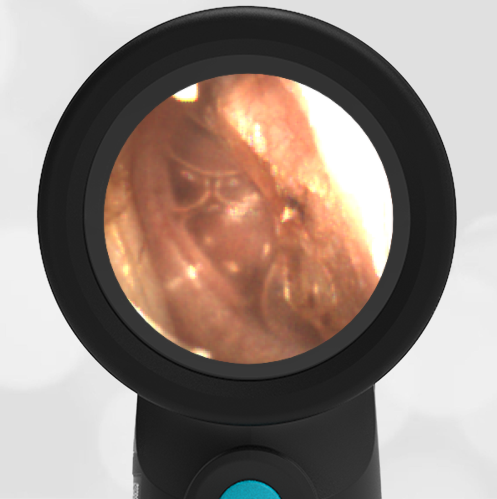
- Severe Bulging
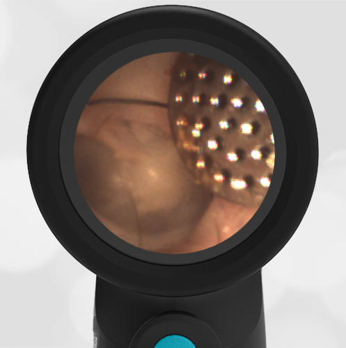
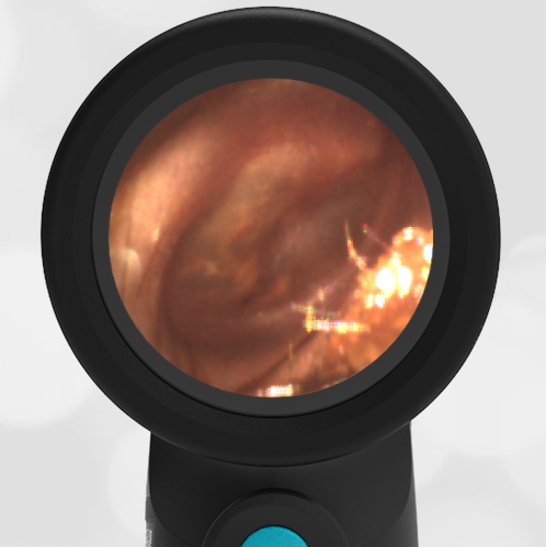
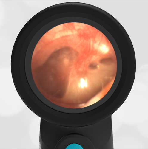
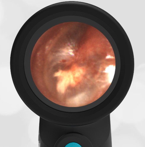
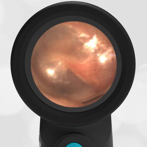
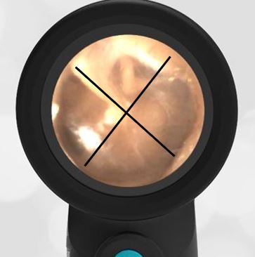
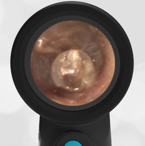
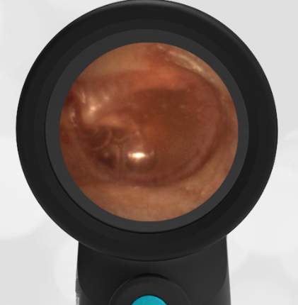
Below is the actual Wispr video from the examination of the 10-month old. The image above was exported by the Wispr from this video. You can see that these views would have been difficult to achieve with a traditional analog otoscope because of canal size and wax.
Video of Acute Otitis Media
If a child experiences recurrent AOM, ventilation tubes may be indicated. AOM and ventilation tubes can both cause sclerosis.
Available at Wispr University is a presentation on normal ear anatomy.