CLINICAL CASES
CLINICAL CASES
Using the Wispr Digital Otoscope


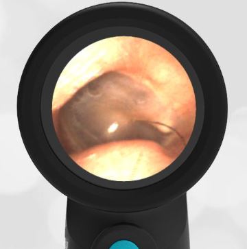
A healthy 26-year-old pediatric resident physician stopped by the WiscMed booth at the Pediatric Academic Society (PAS) conference in Denver in April 2022. A demonstration of the Wispr digital otoscope was performed on her right ear. After seeing the image, the examiner concluded that the resident was a surfer. This was confirmed by the resident.
What unique feature does this image have that supports that conclusion?
Bonus question - what else can you infer about her life from this image?

Exostosis is an abnormal bony growth, in this case in the ear canal. At the tympanic membrane (TM, eardrum) the ear canal is generally “circle-like,” matching the geometry of the TM. You can see that in this photo the bony growth distorts that geometry. Here is an example of a normal ear canal and TM. Exposure to cold water in the ear canal causes this overgrowth. It’s very common to see in surfers, open-water swimmers, and kite surfers. The condition is ongoing, meaning that the more exposure to cold water, the more exostosis increases. In severe cases, bony growth can block the ear canal. This can lead to hearing loss and infection. The condition is benign. As long as there is no hearing loss, pain, or infections, the condition does not need to be addressed. If needed, definitive treatment is via surgery to remove the bony growth. Here is another example of exostosis. What else can you infer from this image? The resident likely has a dog because of the “black hair” sign. It is unknown if the dog is also a surfer.

Here is the complete video of the exam:
Complete exam video

