Otitis Media or Externa – December 21, 2023
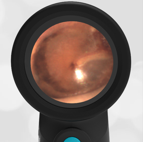
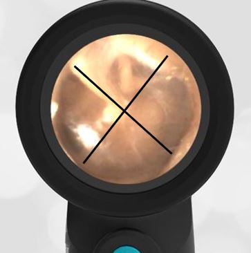
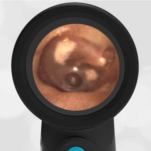
A previously healthy 58-year-old woman contacts her physician-friend with concern for pain just in front of her left ear. She describes the pain as being an ache that seems to localize to the left temporomandibular joint (TMJ). She has no past history of TMJ pain, and she is not diabetic. The discomfort has been getting worse for the past several days. Other than the pain, she has felt well, although she notes that her husband has just come down with Covid. She has tested negative for Covid twice in the past 24 hours. Over-the-counter analgesics have not helped with the discomfort. The following image of her left ear is obtained with a WiscMed Wispr digital otoscope. What is the next step in management?
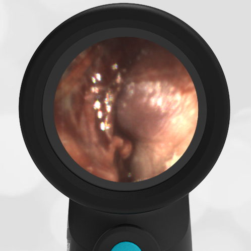
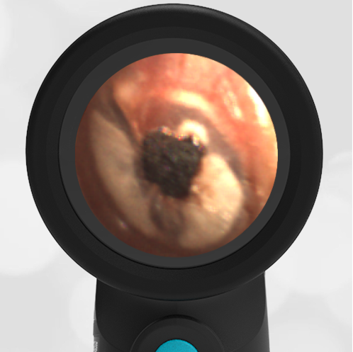
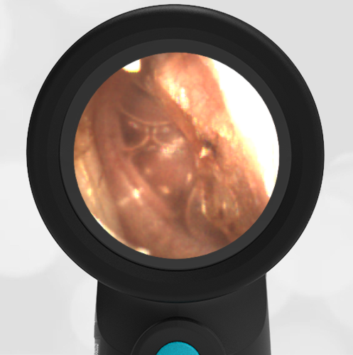
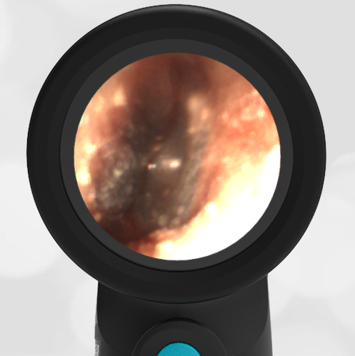
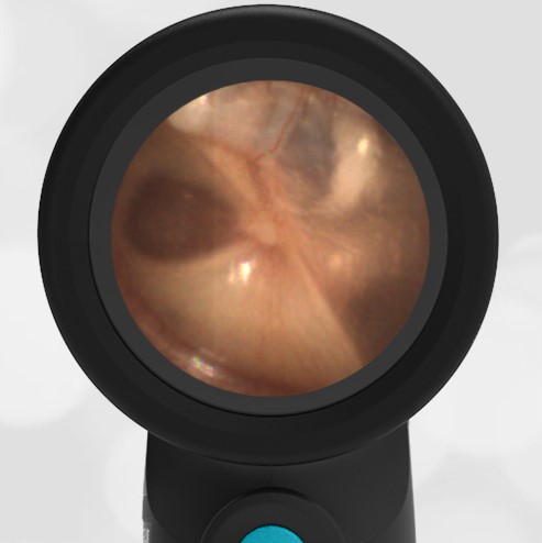
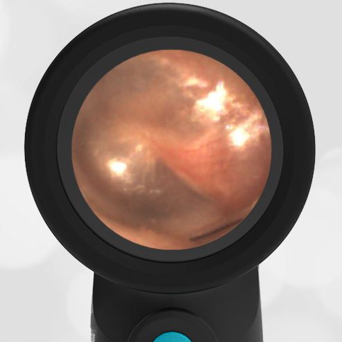
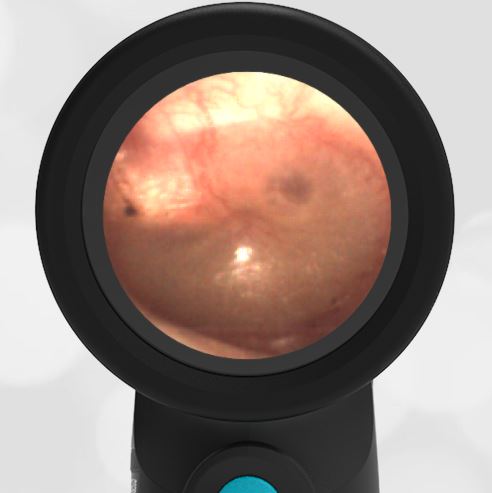
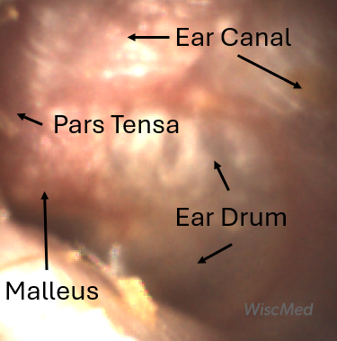 Annotated image
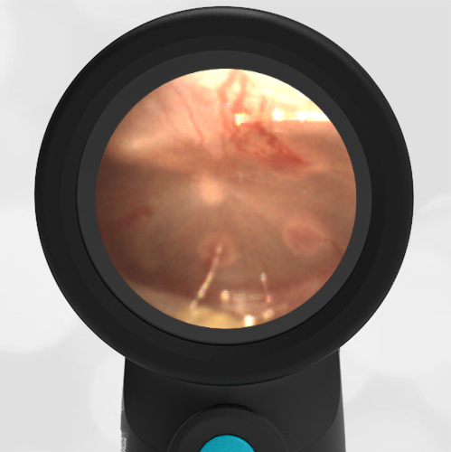
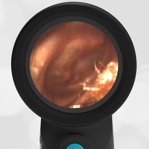
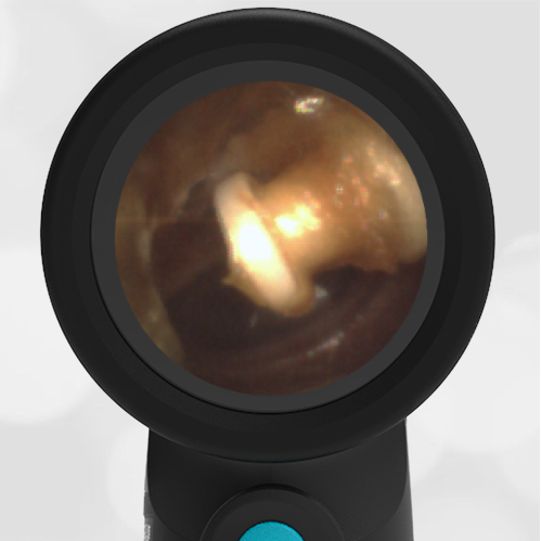
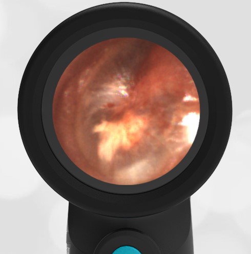
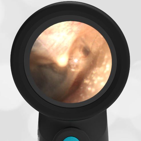
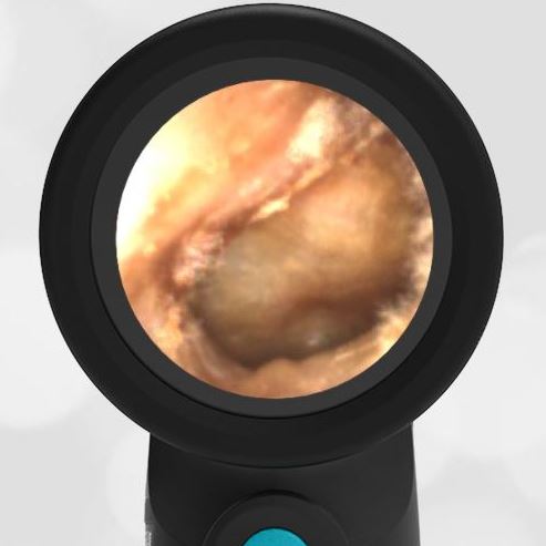
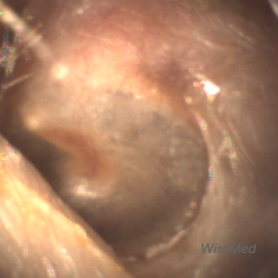
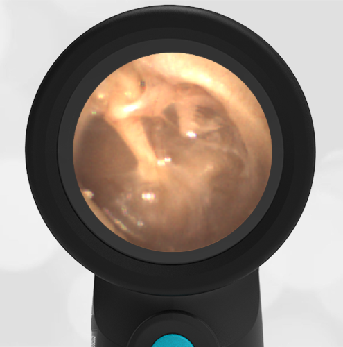
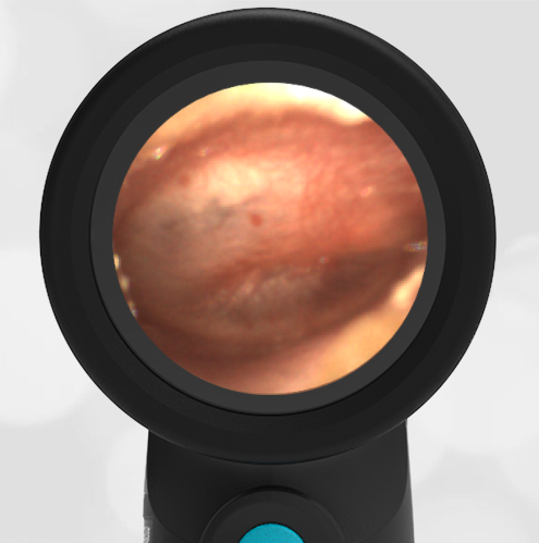
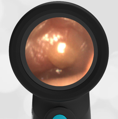
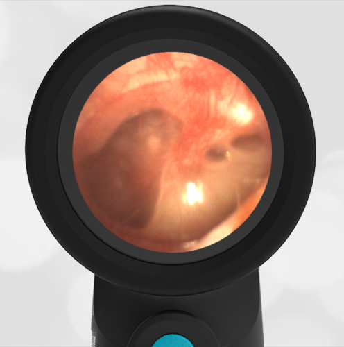
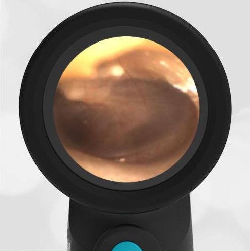
The woman has features of both acute otitis media and otitis externa. The AOM features include bulging of the pars flaccida along with loss of definition of the malleus ossicle. Notably, the pars tensa portion of the ear drum is not bulging and there is no “angry donut” sign. The otitis externa features include inflammation of the external ear canal with erythema. There is no ear canal purulence which is seen with otitis externa.
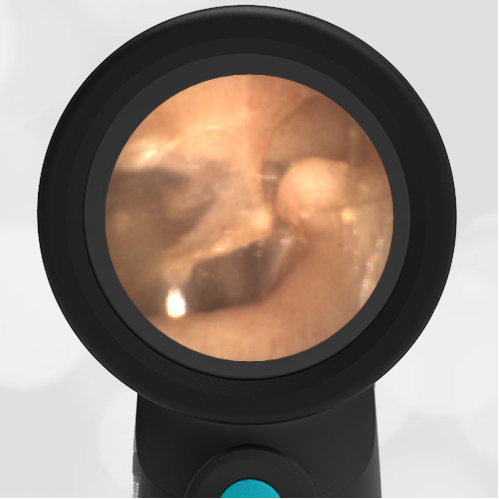
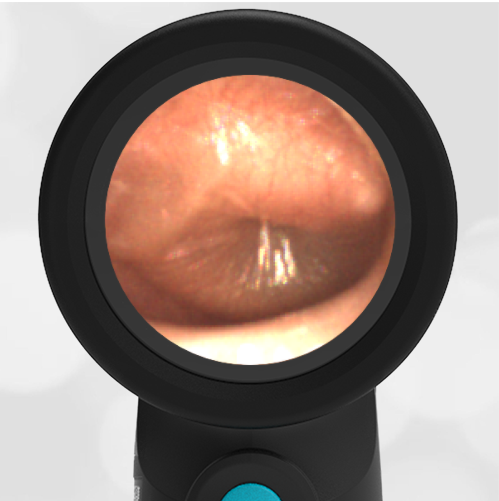
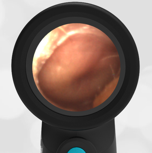
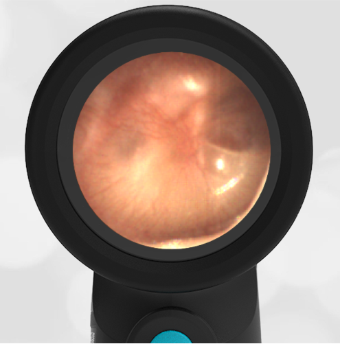
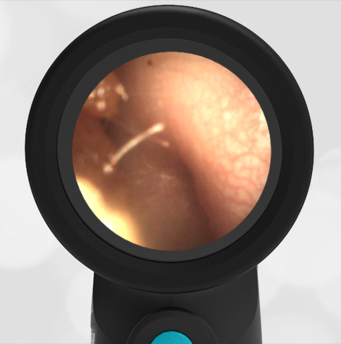
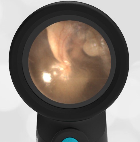
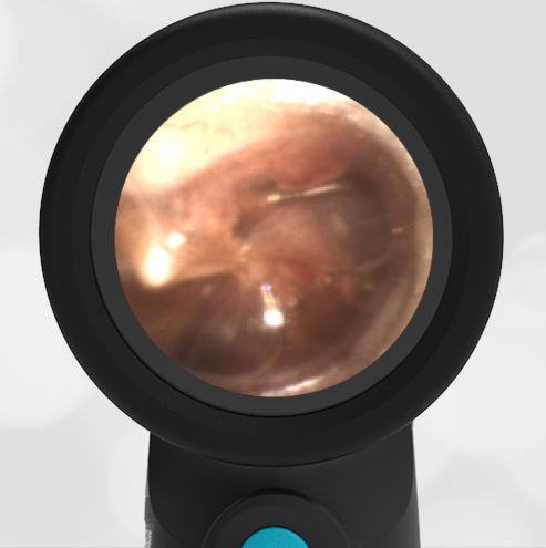
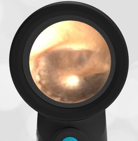
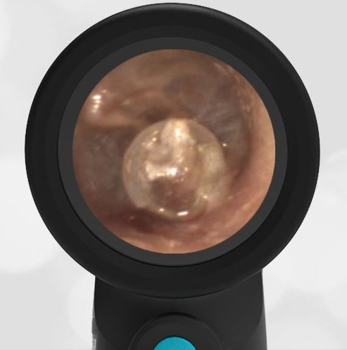
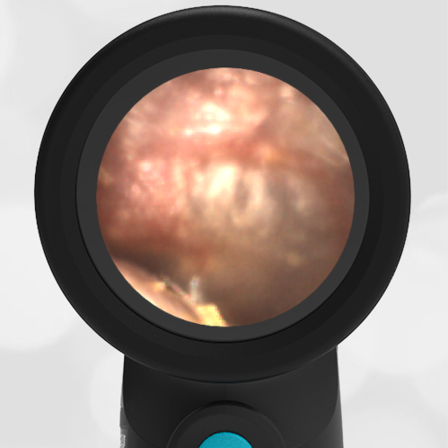
Definitive diagnosis in this case is difficult. The ear is clearly not normal, but it’s not clear if this is a bacterial infection that requires treatment. Because of the discomfort the patient was experiencing along with mild signs of AOM, she was placed on an oral antibiotic. The next day, she reported the symptoms were not better. She was seen in urgent care and placed on an otic antibiotic drop and instructed to continue her oral antibiotic. Over the course of the following week, the discomfort was much reduced. Ten days later, this image of her left ear was obtained. Note that now the malleus is well-defined, the pars flaccida bulging is gone, and there is no erythema.
After 10 days of antibiotics
Here are the complete video exams of the initial presentation and post-treatment.
Pre-Treatment Video Exam
Post-Treatment Video Exam
WiscMed has created a visual diagnosis of ear conditions that may be found here.